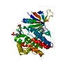
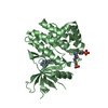
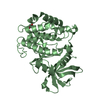
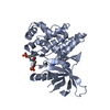
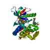
Entry Database : PDB / ID : 6bbuTitle Crystal Structure of JAK1 in complex with compound 25 Tyrosine-protein kinase JAK1 Keywords / / / Function / homology Function Domain/homology Component
/ / / / / / / / / / / / / / / / / / / / / / / / / / / / / / / / / / / / / / / / / / / / / / / / / / / / / / / / / / / / / / / / / / / / / / / / / / / / / / / / / / / / / / / / / / / / / / / / / / / / / / / / / / / / / / / / / / / / / / / / / Biological species Homo sapiens (human)Method / / Resolution : 2.08 Å Authors Han, S. Journal : J. Med. Chem. / Year : 2018Title : Identification of N-{cis-3-[Methyl(7H-pyrrolo[2,3-d]pyrimidin-4-yl)amino]cyclobutyl}propane-1-sulfonamide (PF-04965842): A Selective JAK1 Clinical Candidate for the Treatment of Autoimmune Diseases.Authors: Vazquez, M.L. / Kaila, N. / Strohbach, J.W. / Trzupek, J.D. / Brown, M.F. / Flanagan, M.E. / Mitton-Fry, M.J. / Johnson, T.A. / TenBrink, R.E. / Arnold, E.P. / Basak, A. / Heasley, S.E. / ... Authors : Vazquez, M.L. / Kaila, N. / Strohbach, J.W. / Trzupek, J.D. / Brown, M.F. / Flanagan, M.E. / Mitton-Fry, M.J. / Johnson, T.A. / TenBrink, R.E. / Arnold, E.P. / Basak, A. / Heasley, S.E. / Kwon, S. / Langille, J. / Parikh, M.D. / Griffin, S.H. / Casavant, J.M. / Duclos, B.A. / Fenwick, A.E. / Harris, T.M. / Han, S. / Caspers, N. / Dowty, M.E. / Yang, X. / Banker, M.E. / Hegen, M. / Symanowicz, P.T. / Li, L. / Wang, L. / Lin, T.H. / Jussif, J. / Clark, J.D. / Telliez, J.B. / Robinson, R.P. / Unwalla, R. History Deposition Oct 19, 2017 Deposition site / Processing site Revision 1.0 Jan 17, 2018 Provider / Type Revision 1.1 Feb 21, 2018 Group / Category / citation_authorItem _citation.journal_volume / _citation.page_first ... _citation.journal_volume / _citation.page_first / _citation.page_last / _citation_author.name
Show all Show less
 Movie
Movie Controller


 Open data
 Basic information
 Components
 Keywords
 Kinase /
  Function and homology information
 Authors
 Citation
 Structure visualization
 Downloads & links
 Other downloads
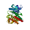
 PDBj
PDBj


 Assembly
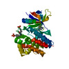


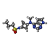
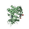
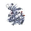
 Mass: 323.414 Da / Num. of mol.: 1 / Source method: obtained synthetically / Formula: C14H21N5O2S / Comment: medication, inhibitor*YM
 Mass: 18.015 Da / Num. of mol.: 127 / Source method: isolated from a natural source / Formula: H2O
 Sample preparation
 / Beamline: 17-ID / Wavelength: 1 Å
 Processing