 Movie
Movie Controller

[English] 日本語
 Yorodumi
- PDB-1bbr: THE STRUCTURE OF RESIDUES 7-16 OF THE A ALPHA CHAIN OF HUMAN FIBR... -
+ Open data

- Basic information
| Entry | Database: PDB / ID: 1bbr | ||||||
|---|---|---|---|---|---|---|---|
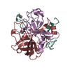
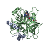
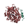
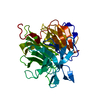
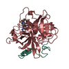
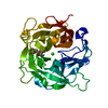
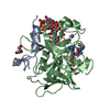
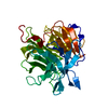
| Title | THE STRUCTURE OF RESIDUES 7-16 OF THE A ALPHA CHAIN OF HUMAN FIBRINOGEN BOUND TO BOVINE THROMBIN AT 2.3 ANGSTROMS RESOLUTION | ||||||
 Components Components |
| ||||||
 Keywords Keywords |  SERINE PROTEASE SERINE PROTEASE | ||||||
| Function / homology |  Function and homology informationblood coagulation, common pathway / induction of bacterial agglutination / fibrinogen complex / Regulation of TLR by endogenous ligand / fibrinogen binding / blood coagulation, fibrin clot formation / platelet alpha granule / MyD88 deficiency (TLR2/4) / positive regulation of heterotypic cell-cell adhesion / IRAK4 deficiency (TLR2/4) ...blood coagulation, common pathway / induction of bacterial agglutination / fibrinogen complex / Regulation of TLR by endogenous ligand / fibrinogen binding / blood coagulation, fibrin clot formation / platelet alpha granule / MyD88 deficiency (TLR2/4) / positive regulation of heterotypic cell-cell adhesion / IRAK4 deficiency (TLR2/4) / MyD88:MAL(TIRAP) cascade initiated on plasma membrane / extracellular matrix structural constituent / plasminogen activation / p130Cas linkage to MAPK signaling for integrins / thrombin / positive regulation of exocytosis / positive regulation of peptide hormone secretion / GRB2:SOS provides linkage to MAPK signaling for Integrins / protein polymerization / Integrin cell surface interactions / positive regulation of blood coagulation / negative regulation of endothelial cell apoptotic process / Common Pathway of Fibrin Clot Formation / positive regulation of substrate adhesion-dependent cell spreading / negative regulation of extrinsic apoptotic signaling pathway via death domain receptors / positive regulation of vasoconstriction / fibrinolysis / Integrin signaling / cell-matrix adhesion / platelet alpha granule lumen / acute-phase response / positive regulation of protein secretion / Post-translational protein phosphorylation / Signaling by high-kinase activity BRAF mutants / MAP2K and MAPK activation / platelet aggregation / platelet activation / response to calcium ion / Signaling by RAF1 mutants / Signaling by moderate kinase activity BRAF mutants / Paradoxical activation of RAF signaling by kinase inactive BRAF / Signaling downstream of RAS mutants / Regulation of Insulin-like Growth Factor (IGF) transport and uptake by Insulin-like Growth Factor Binding Proteins (IGFBPs) / extracellular vesicle / Signaling by BRAF and RAF1 fusions / Platelet degranulation / ER-Phagosome pathway / protein-containing complex assembly / collagen-containing extracellular matrix / blood microparticle / adaptive immune response / positive regulation of ERK1 and ERK2 cascade / Amyloid fiber formation / external side of plasma membrane / endoplasmic reticulum lumen / signaling receptor binding / serine-type endopeptidase activity / innate immune response / calcium ion binding / structural molecule activity / cell surface / endoplasmic reticulum / proteolysis / extracellular space / extracellular exosome / extracellular region / metal ion binding / plasma membrane Function and homology informationblood coagulation, common pathway / induction of bacterial agglutination / fibrinogen complex / Regulation of TLR by endogenous ligand / fibrinogen binding / blood coagulation, fibrin clot formation / platelet alpha granule / MyD88 deficiency (TLR2/4) / positive regulation of heterotypic cell-cell adhesion / IRAK4 deficiency (TLR2/4) ...blood coagulation, common pathway / induction of bacterial agglutination / fibrinogen complex / Regulation of TLR by endogenous ligand / fibrinogen binding / blood coagulation, fibrin clot formation / platelet alpha granule / MyD88 deficiency (TLR2/4) / positive regulation of heterotypic cell-cell adhesion / IRAK4 deficiency (TLR2/4) / MyD88:MAL(TIRAP) cascade initiated on plasma membrane / extracellular matrix structural constituent / plasminogen activation / p130Cas linkage to MAPK signaling for integrins / thrombin / positive regulation of exocytosis / positive regulation of peptide hormone secretion / GRB2:SOS provides linkage to MAPK signaling for Integrins / protein polymerization / Integrin cell surface interactions / positive regulation of blood coagulation / negative regulation of endothelial cell apoptotic process / Common Pathway of Fibrin Clot Formation / positive regulation of substrate adhesion-dependent cell spreading / negative regulation of extrinsic apoptotic signaling pathway via death domain receptors / positive regulation of vasoconstriction / fibrinolysis / Integrin signaling / cell-matrix adhesion / platelet alpha granule lumen / acute-phase response / positive regulation of protein secretion / Post-translational protein phosphorylation / Signaling by high-kinase activity BRAF mutants / MAP2K and MAPK activation / platelet aggregation / platelet activation / response to calcium ion / Signaling by RAF1 mutants / Signaling by moderate kinase activity BRAF mutants / Paradoxical activation of RAF signaling by kinase inactive BRAF / Signaling downstream of RAS mutants / Regulation of Insulin-like Growth Factor (IGF) transport and uptake by Insulin-like Growth Factor Binding Proteins (IGFBPs) / extracellular vesicle / Signaling by BRAF and RAF1 fusions / Platelet degranulation / ER-Phagosome pathway / protein-containing complex assembly / collagen-containing extracellular matrix / blood microparticle / adaptive immune response / positive regulation of ERK1 and ERK2 cascade / Amyloid fiber formation / external side of plasma membrane / endoplasmic reticulum lumen / signaling receptor binding / serine-type endopeptidase activity / innate immune response / calcium ion binding / structural molecule activity / cell surface / endoplasmic reticulum / proteolysis / extracellular space / extracellular exosome / extracellular region / metal ion binding / plasma membraneSimilarity search - Function | ||||||
| Biological species |  Bos taurus (cattle) Bos taurus (cattle) Homo sapiens (human) Homo sapiens (human) | ||||||
| Method | X-RAY DIFFRACTION / Resolution: 2.3 Å | ||||||
 Authors Authors | Martin, P. / Edwards, B. | ||||||
 Citation Citation | Journal: J.Biol.Chem. / Year: 1992 Title: The structure of residues 7-16 of the A alpha-chain of human fibrinogen bound to bovine thrombin at 2.3-A resolution. Authors: Martin, P.D. / Robertson, W. / Turk, D. / Huber, R. / Bode, W. / Edwards, B.F. | ||||||
| History |
|
- Structure visualization
| Structure viewer | Molecule: MolmilJmol/JSmol |
|---|
- Downloads & links
-Download
| PDBx/mmCIF format | 1bbr.cif.gz | 205.8 KB | Display | PDBx/mmCIF format |
|---|---|---|---|---|
| PDB format | pdb1bbr.ent.gz | 168.8 KB | Display | PDB format |
| PDBx/mmJSON format | 1bbr.json.gz | Tree view | PDBx/mmJSON format | |
| Others |  Other downloads Other downloads |
-Validation report
| Arichive directory | https://data.pdbj.org/pub/pdb/validation_reports/bb/1bbrftp://data.pdbj.org/pub/pdb/validation_reports/bb/1bbr | HTTPS FTP |
|---|
-Related structure data
| Similar structure data |
|---|
-Links
 PDBj
PDBj


- Assembly
| Deposited unit | 
| ||||||||
|---|---|---|---|---|---|---|---|---|---|
| 1 | 
| ||||||||
| 2 | 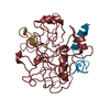
| ||||||||
| 3 | 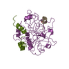
| ||||||||
| 4 | 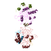
| ||||||||
| Unit cell |
| ||||||||
| Atom site foot note | 1: CIS PROLINE - PRO H 37 / 2: CIS PROLINE - PRO K 37 / 3: CIS PROLINE - PRO N 37 |
-Components
-Protein/peptide , 2 types, 6 molecules LJMFGI
| #1: Protein/peptide | Mass: 5735.240 Da / Num. of mol.: 3 Source method: isolated from a genetically manipulated source Source: (gene. exp.) Bos taurus (cattle) / References: UniProt: P00735, thrombin#4: Protein/peptide | Mass: 1047.144 Da / Num. of mol.: 3 Source method: isolated from a genetically manipulated source Source: (gene. exp.) Homo sapiens (human) / References: UniProt: P02671 |
|---|
-Protein , 3 types, 4 molecules HEKN
| #2: Protein | Mass: 17525.346 Da / Num. of mol.: 1 Source method: isolated from a genetically manipulated source Source: (gene. exp.) Bos taurus (cattle) / References: UniProt: P00735, thrombin |
|---|---|
| #3: Protein | Mass: 12265.071 Da / Num. of mol.: 1 Source method: isolated from a genetically manipulated source Source: (gene. exp.) Bos taurus (cattle) / References: UniProt: P00735, thrombin |
| #5: Protein | Mass: 29772.422 Da / Num. of mol.: 2 Source method: isolated from a genetically manipulated source Source: (gene. exp.) Bos taurus (cattle) / References: UniProt: P00735, thrombin |
-Non-polymers , 1 types, 706 molecules 
| #6: Water | ChemComp-HOH / WaterMass: 18.015 Da / Num. of mol.: 706 / Source method: isolated from a natural source / Formula: H2O |
|---|
-Experimental details
-Experiment
| Experiment | Method: X-RAY DIFFRACTION |
|---|
- Sample preparation
| Crystal | Density Matthews: 3.22 Å3/Da / Density % sol: 61.78 % |
|---|---|
| Crystal grow | *PLUS pH: 8 / Method: vapor diffusion, hanging drop |
| Components of the solutions | *PLUS Conc.: 40 % / Common name: ammonium sulfate |
-Data collection
| Radiation | Scattering type: x-ray |
|---|---|
| Radiation wavelength | Relative weight: 1 |
| Reflection | *PLUS Highest resolution: 2.3 Å / Lowest resolution: 9999 Å / Num. obs: 47459 / % possible obs: 78.9 % / Num. measured all: 176974 / Rmerge(I) obs: 0.075 |
| Reflection shell | *PLUS Highest resolution: 2.3 Å / Lowest resolution: 2.4 Å / % possible obs: 39 % |
- Processing
| Software | Name: PROLSQ / Classification: refinement | |||||||||||||||||||||||||||||||||||||||||||||||||||||||||||||||
|---|---|---|---|---|---|---|---|---|---|---|---|---|---|---|---|---|---|---|---|---|---|---|---|---|---|---|---|---|---|---|---|---|---|---|---|---|---|---|---|---|---|---|---|---|---|---|---|---|---|---|---|---|---|---|---|---|---|---|---|---|---|---|---|---|
| Refinement | Resolution: 2.3→7 Å / Rfactor obs: 0.167 / σ(I): 1 Details: THERE ARE THREE INDEPENDENT COMPLEXES IN THE ASYMMETRIC UNIT. COMPLEX I IS EPSILON THROMBIN (ADDITIONAL CHAIN BREAK BETWEEN RESIDUES 149A AND 149B), AND MOLECULES II AND III ARE ALPHA ...Details: THERE ARE THREE INDEPENDENT COMPLEXES IN THE ASYMMETRIC UNIT. COMPLEX I IS EPSILON THROMBIN (ADDITIONAL CHAIN BREAK BETWEEN RESIDUES 149A AND 149B), AND MOLECULES II AND III ARE ALPHA THROMBIN. FIBRINOPEPTIDE I HAS AN OCCUPANCY THAT MAKES THE AVERAGE B OF ARG-318 ROUGHLY EQUAL TO THAT IN FIBRINOPEPTIDES II AND III. THE DEPOSITORS DO NOT SEE ANY DENSITY AT ALL FOR THROMBIN RESIDUES 1U - 1I, AND POOR DENSITY FOR THE N AND C TERMINI OF THE LIGHT CHAINS, THE C TERMINUS OF THE HEAVY CHAINS, AND THE FIVE RESIDUES BORDERING 149B IN MOLECULES II AND III. EPSILON THROMBIN IN COMPLEX I HAS BEEN ASSIGNED CHAIN INDICATORS *L*, *H*, AND *E*. THE FIBRINOPEPTIDE IN COMPLEX I HAS BEEN ASSIGNED CHAIN INDICATOR *F*. ALPHA THROMBIN IN COMPLEX II HAS BEEN ASSIGNED CHAIN INDICATORS *J* AND *K*. THE FIBRINOPEPTIDE IN COMPLEX II HAS BEEN ASSIGNED CHAIN INDICATOR *G*. ALPHA THROMBIN IN COMPLEX III HAS BEEN ASSIGNED CHAIN INDICATORS *M* AND *N*. THE FIBRINOPEPTIDE IN COMPLEX III HAS BEEN ASSIGNED CHAIN INDICATOR *I*. | |||||||||||||||||||||||||||||||||||||||||||||||||||||||||||||||
| Refinement step | Cycle: LAST / Resolution: 2.3→7 Å
| |||||||||||||||||||||||||||||||||||||||||||||||||||||||||||||||
| Refine LS restraints |
| |||||||||||||||||||||||||||||||||||||||||||||||||||||||||||||||
| Refinement | *PLUS Num. reflection obs: 47459 | |||||||||||||||||||||||||||||||||||||||||||||||||||||||||||||||
| Solvent computation | *PLUS | |||||||||||||||||||||||||||||||||||||||||||||||||||||||||||||||
| Displacement parameters | *PLUS Biso mean: 33.5 Å2 | |||||||||||||||||||||||||||||||||||||||||||||||||||||||||||||||
| Refine LS restraints | *PLUS
|
