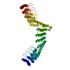
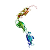
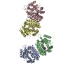
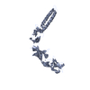
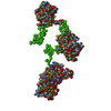
Journal: Structure / Year: 2006 Title: Conformation of polypyrimidine tract binding protein in solution. Authors: Maxim V Petoukhov / Tom P Monie / Frédéric H-T Allain / Stephen Matthews / Stephen Curry / Dmitri I Svergun / Abstract: The polypyrimidine tract binding protein (PTB) is an RNA binding protein that normally functions as a regulator of alternative splicing but can also be recruited to stimulate translation initiation ...The polypyrimidine tract binding protein (PTB) is an RNA binding protein that normally functions as a regulator of alternative splicing but can also be recruited to stimulate translation initiation by certain picornaviruses. High-resolution structures of the four RNA recognition motifs (RRMs) that make up PTB have previously been determined by NMR. Here, we have used small-angle X-ray scattering to determine the low-resolution structure of the entire protein. Scattering patterns from full-length PTB and deletion mutants containing all possible sequential combinations of the RRMs were collected. All constructs were found to be monomeric in solution. Ab initio analysis and rigid-body modeling utilizing the high-resolution models of the RRMs yielded a consistent low-resolution model of the spatial organization of domains in PTB. Domains 3 and 4 were found to be in close contact, whereas domains 2 and especially 1 had loose contacts with the rest of the protein.
In the structure databanks used in Yorodumi, some data are registered as the other names, "COVID-19 virus" and "2019-nCoV". Here are the details of the virus and the list of structure data.
Jan 31, 2019. EMDB accession codes are about to change! (news from PDBe EMDB page)
EMDB accession codes are about to change! (news from PDBe EMDB page)
The allocation of 4 digits for EMDB accession codes will soon come to an end. Whilst these codes will remain in use, new EMDB accession codes will include an additional digit and will expand incrementally as the available range of codes is exhausted. The current 4-digit format prefixed with “EMD-” (i.e. EMD-XXXX) will advance to a 5-digit format (i.e. EMD-XXXXX), and so on. It is currently estimated that the 4-digit codes will be depleted around Spring 2019, at which point the 5-digit format will come into force.
The EM Navigator/Yorodumi systems omit the EMD- prefix.
Related info.:Q: What is EMD? / ID/Accession-code notation in Yorodumi/EM Navigator
Yorodumi is a browser for structure data from EMDB, PDB, SASBDB, etc.
This page is also the successor to EM Navigator detail page, and also detail information page/front-end page for Omokage search.
The word "yorodu" (or yorozu) is an old Japanese word meaning "ten thousand". "mi" (miru) is to see.
Related info.:EMDB / PDB / SASBDB / Comparison of 3 databanks / Yorodumi Search / Aug 31, 2016. New EM Navigator & Yorodumi / Yorodumi Papers / Jmol/JSmol / Function and homology information / Changes in new EM Navigator and Yorodumi
 Movie
Movie Controller

 Open data
 Basic information
 Sample
 Function and homology information
 Homo sapiens (human)
 Citation
 Contact author
 Structure visualization
 Downloads & links
 SASDAR4


 Search similar-shape structures of this assembly by Omokage search (details)
 DORIS III X33


