 Movie
Movie Controller

[English] 日本語
 Yorodumi
- SASDGD5: The PDZ1-2 domain of postsynaptic density protein 95 (PSD-95) bou... -
+ Open data

- Basic information
| Entry | Database: SASBDB / ID: SASDGD5 |
|---|---|
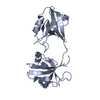
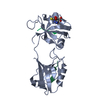
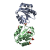
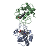
 Sample Sample | The PDZ1-2 domain of postsynaptic density protein 95 (PSD-95) bound to RRESEI peptide (Paused SEC)
|
| Function / homology |  Function and homology information Function and homology informationLGI-ADAM interactions / P2Y1 nucleotide receptor binding / beta-1 adrenergic receptor binding / neuroligin family protein binding / regulation of grooming behavior / NrCAM interactions / synaptic vesicle maturation / positive regulation of neuron projection arborization / receptor localization to synapse / establishment or maintenance of epithelial cell apical/basal polarity ...LGI-ADAM interactions / P2Y1 nucleotide receptor binding / beta-1 adrenergic receptor binding / neuroligin family protein binding / regulation of grooming behavior / NrCAM interactions / synaptic vesicle maturation / positive regulation of neuron projection arborization / receptor localization to synapse / establishment or maintenance of epithelial cell apical/basal polarity / Synaptic adhesion-like molecules / cerebellar mossy fiber / protein localization to synapse / Trafficking of AMPA receptors / neuron spine / dendritic spine morphogenesis / negative regulation of receptor internalization / juxtaparanode region of axon / vocalization behavior / acetylcholine receptor binding / RHO GTPases activate CIT / cellular response to potassium ion / Assembly and cell surface presentation of NMDA receptors / NMDA selective glutamate receptor signaling pathway / Neurexins and neuroligins / neuromuscular process controlling balance / Activation of Ca-permeable Kainate Receptor / neurotransmitter receptor localization to postsynaptic specialization membrane / neuron projection terminus / cortical cytoskeleton / Negative regulation of NMDA receptor-mediated neuronal transmission / Unblocking of NMDA receptors, glutamate binding and activation / AMPA glutamate receptor clustering / Signaling by ERBB4 / locomotory exploration behavior / AMPA glutamate receptor complex / Long-term potentiation / social behavior / D1 dopamine receptor binding / positive regulation of synaptic transmission / regulation of postsynaptic membrane neurotransmitter receptor levels / excitatory synapse / ionotropic glutamate receptor binding / dendrite cytoplasm / Ras activation upon Ca2+ influx through NMDA receptor / positive regulation of excitatory postsynaptic potential / learning / synaptic membrane / adherens junction / PDZ domain binding / neuromuscular junction / establishment of protein localization / cell-cell adhesion / regulation of long-term neuronal synaptic plasticity / postsynaptic density membrane / kinase binding / endocytic vesicle membrane / synaptic vesicle / cell junction / nervous system development / positive regulation of cytosolic calcium ion concentration / RAF/MAP kinase cascade / protein-containing complex assembly / scaffold protein binding / protein phosphatase binding / dendritic spine / chemical synaptic transmission / protein-macromolecule adaptor activity / postsynaptic membrane / neuron projection / postsynaptic density / synapse / protein kinase binding / protein-containing complex binding / glutamatergic synapse / endoplasmic reticulum / signal transduction / plasma membrane / cytoplasm / cytosol Similarity search - Function |
| Biological species |  Homo sapiens (human) Homo sapiens (human) |
 Citation Citation | Date: 2019 Sep 19 Title: How the dual PDZ domain from Postsynaptic density protein 95 clusters ion channels and receptors Authors: Rodzli N / Lockhart-Cairns M / Levy C / Chipperfield J / Bird L / Baldock C |
 Contact author Contact author |
|
- Structure visualization
| Structure viewer | Molecule: MolmilJmol/JSmol |
|---|
- Downloads & links
-Data source
| SASBDB page |  SASDGD5 SASDGD5 |
|---|
-Related structure data
| Related structure data | C: citing same article ( |
|---|---|
| Similar structure data |


-External links
| Related items in Molecule of the Month |
|---|


-Models


-Sample
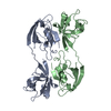
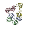
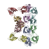
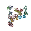
| Sample | Name: The PDZ1-2 domain of postsynaptic density protein 95 (PSD-95) bound to RRESEI peptide (Paused SEC) Specimen concentration: 15 mg/ml |
|---|---|
| Buffer | Name: 20 mM TRIS/HCl, 150 mM NaCl + 10 mM RRESEI / pH: 8.5 / Comment: standard buffer plus 10mM ligand peptide |
| Entity #1889 | Name: PDZ1-2 / Type: protein Description: PDZ1-2 fragment of PSD-95/Disks large homolog 4 Formula weight: 20.8 / Num. of mol.: 1 / Source: Homo sapiens / References: UniProt: P78352 Sequence: GPGTEGEMEY EEITLERGNS GLGFSIAGGT DNPHIGDDPS IFITKIIPGG AAAQDGRLRV NDSILFVNEV DVREVTHSAA VEALKEAGSI VRLYVMRRKP PAEKVMEIKL IKGPKGLGFS IAGGVGNQHI PGDNSIYVTK IIEGGAAHKD GRLQIGDKIL AVNSVGLEDV ...Sequence: GPGTEGEMEY EEITLERGNS GLGFSIAGGT DNPHIGDDPS IFITKIIPGG AAAQDGRLRV NDSILFVNEV DVREVTHSAA VEALKEAGSI VRLYVMRRKP PAEKVMEIKL IKGPKGLGFS IAGGVGNQHI PGDNSIYVTK IIEGGAAHKD GRLQIGDKIL AVNSVGLEDV MHEDAVAALK NTYDVVYLKV AKPSNA |
-Experimental information
| Beam | Instrument name: Diamond Light Source B21 / City: Didcot / 国: UK  / Shape: 1 x 5 mm / Type of source: X-ray synchrotron / Wavelength: 0.1 Å / Dist. spec. to detc.: 3.9 mm / Shape: 1 x 5 mm / Type of source: X-ray synchrotron / Wavelength: 0.1 Å / Dist. spec. to detc.: 3.9 mm | ||||||||||||||||||||||||||||||
|---|---|---|---|---|---|---|---|---|---|---|---|---|---|---|---|---|---|---|---|---|---|---|---|---|---|---|---|---|---|---|---|
| Detector | Name: Pilatus 2M | ||||||||||||||||||||||||||||||
| Scan |  Measurement date: Jan 26, 2016 / Exposure time: 10 sec. / Number of frames: 69 / Unit: 1/A /
| ||||||||||||||||||||||||||||||
| Distance distribution function P(R) | 
| ||||||||||||||||||||||||||||||
| Result |  Comments: Scattering data are fitted using the ATSAS OLIGOMER program using a suite of models. The ATSAS program FFMAKER was used to generate form factors for each model in the suite. Each model is ...Comments: Scattering data are fitted using the ATSAS OLIGOMER program using a suite of models. The ATSAS program FFMAKER was used to generate form factors for each model in the suite. Each model is assigned a volume fraction to fit the observed scattering profile. 3 monomer models are included, the first is a compact conformation of PDZ1-2 similar to PDB entries 6spv/6spz; the other two are domain models obtained from a representative run of the ATSAS EOM program with data projected to infinite dilution. Multimeric models are drawn from a "clustering Spacegroup", unit cell 14.8nm symmetry I2(1)3, and consist of identical copies of an extended conformation of PDZ1-2 (similar to PDB entry 3zrt) assembled by symmetry operations. In the order of deposition: Model number; Stoichiometry; MW (kDa); source; Volume fraction. 1; 1; 21; Crystal Structure; 0.381. 2; 1; 21; EOM; 0.091. 3; 1; 21; EOM; 0.470. 4; 2; 42; clustering Spacegroup; 0.042. 5; 4; 84; clustering Spacegroup; 0.015. 6; 8; 168; clustering Spacegroup; 0.000. 7; 12, 252; clustering Spacegroup; 0.001. 8; 16, 336, clustering Spacegroup; 0.000. 9; 20, 420, clustering Spacegroup; 0.000. 10; 24, 504, clustering Spacegroup; 0.001.
|
