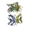
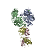
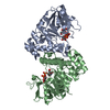
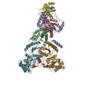
+ Open data
- Basic information
| Entry | Database: SASBDB / ID: SASDA48 |
|---|---|
 Sample Sample | transglutaminase2:anti-transglutaminase2 FAB1 antibody complex
|
| Biological species |  Homo sapiens (human) Homo sapiens (human) |
 Citation Citation |  Journal: J Biol Chem / Year: 2015 Journal: J Biol Chem / Year: 2015Title: Structural Basis for Antigen Recognition by Transglutaminase 2-specific Autoantibodies in Celiac Disease. Authors: Xi Chen / Kathrin Hnida / Melissa Ann Graewert / Jan Terje Andersen / Rasmus Iversen / Anne Tuukkanen / Dmitri Svergun / Ludvig M Sollid /   Abstract: Antibodies to the autoantigen transglutaminase 2 (TG2) are a hallmark of celiac disease. We have studied the interaction between TG2 and an anti-TG2 antibody (679-14-E06) derived from a single gut ...Antibodies to the autoantigen transglutaminase 2 (TG2) are a hallmark of celiac disease. We have studied the interaction between TG2 and an anti-TG2 antibody (679-14-E06) derived from a single gut IgA plasma cell of a celiac disease patient. The antibody recognizes one of four identified epitopes targeted by antibodies of plasma cells of the disease lesion. The binding interface was identified by small angle x-ray scattering, ab initio and rigid body modeling using the known crystal structure of TG2 and the crystal structure of the antibody Fab fragment, which was solved at 2.4 Å resolution. The result was confirmed by testing binding of the antibody to TG2 mutants by ELISA and surface plasmon resonance. TG2 residues Arg-116 and His-134 were identified to be critical for binding of 679-14-E06 as well as other epitope 1 antibodies. In contrast, antibodies directed toward the two other main epitopes (epitopes 2 and 3) were not affected by these mutations. Molecular dynamics simulations suggest interactions of 679-14-E06 with the N-terminal domain of TG2 via the CDR2 and CDR3 loops of the heavy chain and the CDR2 loop of the light chain. In addition there were contacts of the framework 3 region of the heavy chain with the catalytic domain of TG2. The results provide an explanation for the biased usage of certain heavy and light chain gene segments by epitope 1-specific antibodies in celiac disease. |
 Contact author Contact author |
|
- Structure visualization
| Structure viewer | Molecule:  Molmil Molmil Jmol/JSmol Jmol/JSmol |
|---|
- Downloads & links
-Models
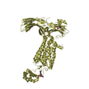
| Model #308 | 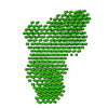 Type: dummy / Software: DAMMIN / Radius of dummy atoms: 3.50 A / Chi-square value: 0.724201 / P-value: 0.157000  Search similar-shape structures of this assembly by Omokage search (details) Search similar-shape structures of this assembly by Omokage search (details) |
|---|---|
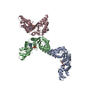
| Model #309 |  Type: atomic / Software: SASREF / Radius of dummy atoms: 1.90 A / Chi-square value: 0.66 / P-value: 0.053000  Search similar-shape structures of this assembly by Omokage search (details) Search similar-shape structures of this assembly by Omokage search (details) |
- Sample
 Sample Sample | Name: transglutaminase2:anti-transglutaminase2 FAB1 antibody complex Specimen concentration: 9 mg/ml / Entity id: 175 / 176 |
|---|---|
| Buffer | Name: 20 mM Tris 150mM NaCl 1mM EDTA / Concentration: 20.00 mM / pH: 7.2 / Composition: 150mM NaCl, 1mM EDTA |
| Entity #175 | Type: protein / Description: anti-TG2 antibody (679 14 E06) / Formula weight: 47.639 / Num. of mol.: 1 Sequence: EVQLVQSGAE VKKPGESLKI SCKGSGYSFT SYWIGWVRQM PGKGLEWMGI IYPGDSDTRY SPSFQGQVTI SADKSISTAY LQWSSLKASD TAMYYCARPH YYDSLDAFDI WGQGTMVTVS SASTKGPSVF PLAPSSKSTS GGTAALGCLV KDYFPEPVTV SWNSGALTSG ...Sequence: EVQLVQSGAE VKKPGESLKI SCKGSGYSFT SYWIGWVRQM PGKGLEWMGI IYPGDSDTRY SPSFQGQVTI SADKSISTAY LQWSSLKASD TAMYYCARPH YYDSLDAFDI WGQGTMVTVS SASTKGPSVF PLAPSSKSTS GGTAALGCLV KDYFPEPVTV SWNSGALTSG VHTFPAVLQS SGLYSLSSVV TVPSSSLGTQ TYICNVNHKP SNTKVDKRVE PKSCDIQMTQ SPSTLSASVG DRVTITCRAS QSISSWLAWY QQRPGKAPKL LIYKASSLES GVPSRFSGSG SGTEFTLTIS SLQPDDFATY YCQHYNSYSP GYTFGQGTKV EIKRTVAAPS VFIFPPSDEQ LKSGTASVVC LLNNFYPREA KVQWKVDNAL QSGNSQESVT EQDSKDSTYS LSSTLTLSKA DYEKHKVYAC EVTHQGLSSP VTKSFNRGEC |
| Entity #176 | Name: TGA / Type: protein / Description: transglutaminase 2 / Formula weight: 79.491 / Num. of mol.: 1 / Source: Homo sapiens Sequence: MGSSHHHHHH SSGLVPRGSH maeelvlerc dleletngrd hhtadlcrek lvvrrgqpfw ltlhfegrny easvdsltfs vvtgpapsqe agtkarfplr daveegdwta tvvdqqdctl slqlttpana piglyrlsle astgyqgssf vlghfillfn awcpadavyl ...Sequence: MGSSHHHHHH SSGLVPRGSH maeelvlerc dleletngrd hhtadlcrek lvvrrgqpfw ltlhfegrny easvdsltfs vvtgpapsqe agtkarfplr daveegdwta tvvdqqdctl slqlttpana piglyrlsle astgyqgssf vlghfillfn awcpadavyl dseeerqeyv ltqqgfiyqg sakfiknipw nfgqfedgil diclilldvn pkflknagrd csrrsspvyv grvvsgmvnc nddqgvllgr wdnnygdgvs pmswigsvdi lrrwknhgcq rvkygqcwvf aavactvlrc lgiptrvvtn ynsahdqnsn llieyfrnef geiqgdksem iwnfhcwves wmtrpdlqpg yegwqaldpt pqeksegtyc cgpvpvraik egdlstkyda pfvfaevnad vvdwiqqddg svhksinrsl ivglkistks vgrderedit htykypegss eereaftran hlnklaekee tgmamrirvg qsmnmgsdfd vfahitnnta eeyvcrlllc artvsyngil gpecgtkyll nlnlepfsek svplcilyek yrdcltesnl ikvrallvep vinsyllaer dlylenpeik irilgepkqk rklvaevslq nplpvalegc tftvegaglt eeqktveipd pveageevkv rmdllplhmg lhklvvnfes dklkavkgfr nviigpa |
-Experimental information
| Beam | Instrument name: PETRA III P12 / City: Hamburg / 国: Germany  / Type of source: X-ray synchrotron / Wavelength: 0.12 Å / Dist. spec. to detc.: 3.1 mm / Type of source: X-ray synchrotron / Wavelength: 0.12 Å / Dist. spec. to detc.: 3.1 mm | |||||||||||||||||||||||||||||||||||||||
|---|---|---|---|---|---|---|---|---|---|---|---|---|---|---|---|---|---|---|---|---|---|---|---|---|---|---|---|---|---|---|---|---|---|---|---|---|---|---|---|---|
| Detector | Name: Pilatus 2M | |||||||||||||||||||||||||||||||||||||||
| Scan |
| |||||||||||||||||||||||||||||||||||||||
| Distance distribution function P(R) |
| |||||||||||||||||||||||||||||||||||||||
| Result | Comments: Structural basis for antigen recognition by transglutaminase 2-specific autoantibodies in celiac disease. SAXS profile of the complex formed between transglutaminase2 bound to anti- ...Comments: Structural basis for antigen recognition by transglutaminase 2-specific autoantibodies in celiac disease. SAXS profile of the complex formed between transglutaminase2 bound to anti-transglutaminase2 antibody (679 14 E06) FAB1.
|
 Movie
Movie Controller


 SASDA48