 Movie
Movie Controller

[English] 日本語
 Yorodumi
- PDB-9znd: Structure of rabbit RyR1 complexed with FKBP12.6 and calmodulin i... -
+ Open data

- Basic information
| Entry | Database: PDB / ID: 9znd | |||||||||
|---|---|---|---|---|---|---|---|---|---|---|
| Title | Structure of rabbit RyR1 complexed with FKBP12.6 and calmodulin in the presence of dantrolene, ATP, 4-chloro-m-cresol, caffeine, and Mg2+ | |||||||||
 Components Components |
| |||||||||
 Keywords Keywords | MEMBRANE PROTEIN / sarcoplasmic reticulum / intracellular calcium channel / excitation-contraction coupling / complex | |||||||||
| Function / homology |  Function and homology information Function and homology informationnegative regulation of calcium-mediated signaling / ATP-gated ion channel activity / negative regulation of release of sequestered calcium ion into cytosol / response to redox state / ryanodine-sensitive calcium-release channel activity / terminal cisterna / CaM pathway / Cam-PDE 1 activation / negative regulation of heart rate / Sodium/Calcium exchangers ...negative regulation of calcium-mediated signaling / ATP-gated ion channel activity / negative regulation of release of sequestered calcium ion into cytosol / response to redox state / ryanodine-sensitive calcium-release channel activity / terminal cisterna / CaM pathway / Cam-PDE 1 activation / negative regulation of heart rate / Sodium/Calcium exchangers / release of sequestered calcium ion into cytosol by sarcoplasmic reticulum / ossification involved in bone maturation / Calmodulin induced events / ryanodine receptor complex / Reduction of cytosolic Ca++ levels / Activation of Ca-permeable Kainate Receptor / CREB1 phosphorylation through the activation of CaMKII/CaMKK/CaMKIV cascasde / cellular response to caffeine / Loss of phosphorylation of MECP2 at T308 / skin development / CREB1 phosphorylation through the activation of Adenylate Cyclase / PKA activation / CaMK IV-mediated phosphorylation of CREB / FK506 binding / 'de novo' protein folding / CASP4 inflammasome assembly / Glycogen breakdown (glycogenolysis) / negative regulation of ryanodine-sensitive calcium-release channel activity / Activation of RAC1 downstream of NMDARs / organelle localization by membrane tethering / CLEC7A (Dectin-1) induces NFAT activation / : / organelle membrane / negative regulation of high voltage-gated calcium channel activity / autophagosome membrane docking / negative regulation of calcium ion export across plasma membrane / regulation of cardiac muscle cell action potential / presynaptic endocytosis / outflow tract morphogenesis / Synthesis of IP3 and IP4 in the cytosol / Phase 0 - rapid depolarisation / Negative regulation of NMDA receptor-mediated neuronal transmission / smooth endoplasmic reticulum / Unblocking of NMDA receptors, glutamate binding and activation / intracellularly gated calcium channel activity / calcineurin-mediated signaling / RHO GTPases activate PAKs / regulation of cell communication by electrical coupling involved in cardiac conduction / Uptake and function of anthrax toxins / Ion transport by P-type ATPases / protein phosphatase activator activity / Long-term potentiation / Calcineurin activates NFAT / regulation of ryanodine-sensitive calcium-release channel activity / DARPP-32 events / Regulation of MECP2 expression and activity / Smooth Muscle Contraction / detection of calcium ion / regulation of cardiac muscle contraction / cellular response to interferon-beta / toxic substance binding / presynaptic cytosol / RHO GTPases activate IQGAPs / catalytic complex / calcium channel inhibitor activity / skeletal muscle fiber development / voltage-gated calcium channel activity / release of sequestered calcium ion into cytosol / eNOS activation / regulation of release of sequestered calcium ion into cytosol by sarcoplasmic reticulum / Tetrahydrobiopterin (BH4) synthesis, recycling, salvage and regulation / Activation of AMPK downstream of NMDARs / Ion homeostasis / regulation of heart rate / Protein methylation / regulation of cardiac muscle contraction by regulation of the release of sequestered calcium ion / titin binding / calcium channel complex / cellular response to calcium ion / voltage-gated potassium channel complex / FCERI mediated Ca+2 mobilization / muscle contraction / substantia nigra development / sarcoplasmic reticulum membrane / FCGR3A-mediated IL10 synthesis / protein serine/threonine kinase activator activity / sperm midpiece / Antigen activates B Cell Receptor (BCR) leading to generation of second messengers / positive regulation of receptor signaling pathway via JAK-STAT / calyx of Held / Ras activation upon Ca2+ influx through NMDA receptor / adenylate cyclase activator activity / regulation of cytokinesis / VEGFR2 mediated cell proliferation / calcium-mediated signaling / spindle microtubule / VEGFR2 mediated vascular permeability / sarcomere / peptidylprolyl isomerase / sarcoplasmic reticulum Similarity search - Function | |||||||||
| Biological species |  Homo sapiens (human) Homo sapiens (human)  Oryctolagus cuniculus (rabbit) Oryctolagus cuniculus (rabbit) | |||||||||
| Method | ELECTRON MICROSCOPY / single particle reconstruction / cryo EM / Resolution: 3.09 Å | |||||||||
 Authors Authors | Kim, K. / Clarke, O.B. | |||||||||
| Funding support |  United States, 1items United States, 1items
| |||||||||
 Citation Citation | Journal: Nat Commun / Year: 2026 Title: Structural identification of the RY12 domain of RyR1 as an ADP sensor and the target of the malignant hyperthermia therapeutic dantrolene Authors: Kim, K. / Li, H. / Yuan, Q. / Melville, Z. / Zalk, R. / des Georges, A. / Frank, J. / Hendrickson, W.A. / Marks, A.R. / Clarke, O.B. | |||||||||
| History |
|
- Structure visualization
| Structure viewer | Molecule: MolmilJmol/JSmol |
|---|
- Downloads & links
-Download
| PDBx/mmCIF format | 9znd.cif.gz | 3.4 MB | Display | PDBx/mmCIF format |
|---|---|---|---|---|
| PDB format | pdb9znd.ent.gz | Display | PDB format | |
| PDBx/mmJSON format | 9znd.json.gz | Tree view | PDBx/mmJSON format | |
| Others |  Other downloads Other downloads |
-Validation report
| Arichive directory | https://data.pdbj.org/pub/pdb/validation_reports/zn/9zndftp://data.pdbj.org/pub/pdb/validation_reports/zn/9znd | HTTPS FTP |
|---|
-Related structure data
| Related structure data |  74445MC  9zk8C  9zk9C  9zkaC  9zkcC  9zkdC  9zkeC  9zl7C  9zlcC  9zlfC  9zlhC  9zn6C  9zn7C  9zn8C  9zn9C  9znaC C: citing same article ( M: map data used to model this data |
|---|---|
| Similar structure data |


-Links
 PDBj
PDBj











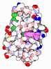
- Assembly
| Deposited unit | 
|
|---|---|
| 1 |
|
-Components
-Protein , 3 types, 12 molecules EFGHIJKLDBAC
| #1: Protein | Mass: 11667.305 Da / Num. of mol.: 4 Source method: isolated from a genetically manipulated source Source: (gene. exp.) Homo sapiens (human) / Gene: FKBP1B, FKBP12.6, FKBP1L, FKBP9, OTK4 / Production host:   Escherichia coli (E. coli) / References: UniProt: P68106, peptidylprolyl isomerase Escherichia coli (E. coli) / References: UniProt: P68106, peptidylprolyl isomerase#2: Protein | Mass: 16790.434 Da / Num. of mol.: 4 Source method: isolated from a genetically manipulated source Source: (gene. exp.) Homo sapiens (human) / Gene: CALM1, CALM, CAM, CAM1 / Production host: Escherichia coli (E. coli) / References: UniProt: P0DP23#3: Protein | Mass: 565908.625 Da / Num. of mol.: 4 / Source method: isolated from a natural source / Source: (natural) Oryctolagus cuniculus (rabbit) / References: UniProt: P11716 |
|---|
-Non-polymers , 8 types, 1734 molecules 

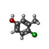
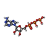
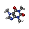

| #4: Chemical | ChemComp-CA / 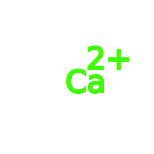 Mass: 40.078 Da / Num. of mol.: 4 / Source method: obtained synthetically / Formula: Ca Mass: 40.078 Da / Num. of mol.: 4 / Source method: obtained synthetically / Formula: Ca#5: Chemical | ChemComp-ZN / 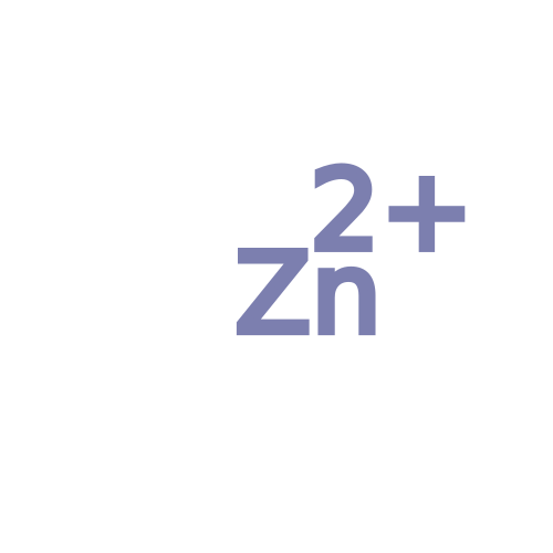 Mass: 65.409 Da / Num. of mol.: 4 / Source method: obtained synthetically / Formula: Zn Mass: 65.409 Da / Num. of mol.: 4 / Source method: obtained synthetically / Formula: Zn#6: Chemical | ChemComp-U1C / Mass: 314.253 Da / Num. of mol.: 4 / Source method: obtained synthetically / Formula: C14H10N4O5 / Feature type: SUBJECT OF INVESTIGATION #7: Chemical | ChemComp-43M / 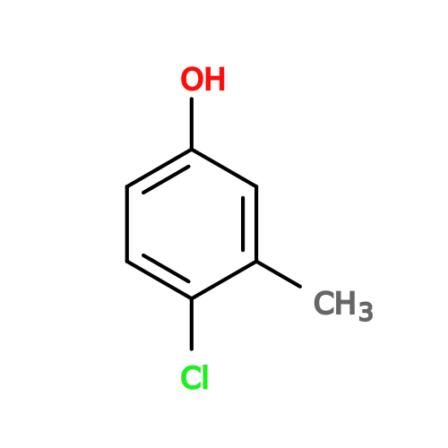 Mass: 142.583 Da / Num. of mol.: 12 / Source method: obtained synthetically / Formula: C7H7ClO / Feature type: SUBJECT OF INVESTIGATION Mass: 142.583 Da / Num. of mol.: 12 / Source method: obtained synthetically / Formula: C7H7ClO / Feature type: SUBJECT OF INVESTIGATION#8: Chemical | ChemComp-ATP / 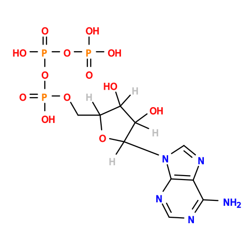 Mass: 507.181 Da / Num. of mol.: 8 / Source method: obtained synthetically / Formula: C10H16N5O13P3 / Feature type: SUBJECT OF INVESTIGATION / Comment: ATP, energy-carrying molecule*YM Mass: 507.181 Da / Num. of mol.: 8 / Source method: obtained synthetically / Formula: C10H16N5O13P3 / Feature type: SUBJECT OF INVESTIGATION / Comment: ATP, energy-carrying molecule*YM#9: Chemical | ChemComp-CFF / 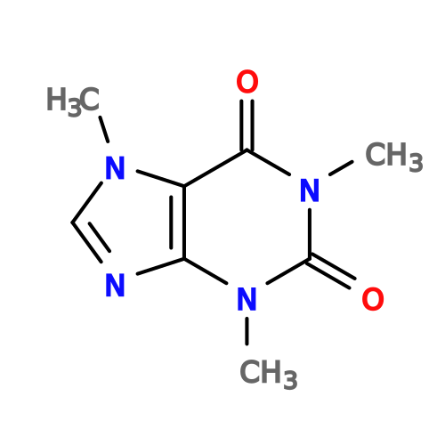 Mass: 194.191 Da / Num. of mol.: 4 / Source method: obtained synthetically / Formula: C8H10N4O2 / Comment: medication*YM Mass: 194.191 Da / Num. of mol.: 4 / Source method: obtained synthetically / Formula: C8H10N4O2 / Comment: medication*YM#10: Chemical | ChemComp-CPS /  Mass: 614.877 Da / Num. of mol.: 4 / Source method: obtained synthetically / Formula: C32H58N2O7S / Comment: detergent*YM Mass: 614.877 Da / Num. of mol.: 4 / Source method: obtained synthetically / Formula: C32H58N2O7S / Comment: detergent*YM#11: Water | ChemComp-HOH / | Mass: 18.015 Da / Num. of mol.: 1694 / Source method: isolated from a natural source / Formula: H2O |
|---|
-Details
| Has ligand of interest | Y |
|---|---|
| Has protein modification | Y |
-Experimental details
-Experiment
| Experiment | Method: ELECTRON MICROSCOPY |
|---|---|
| EM experiment | Aggregation state: PARTICLE / 3D reconstruction method: single particle reconstruction |
- Sample preparation
| Component | Name: RyR1 complexed with FKBP12.6 and calmodulin / Type: COMPLEX / Entity ID: #3, #1-#2 / Source: MULTIPLE SOURCES |
|---|---|
| Buffer solution | pH: 7.5 |
| Specimen | Embedding applied: NO / Shadowing applied: NO / Staining applied: NO / Vitrification applied: YES |
| Vitrification | Cryogen name: ETHANE |
- Electron microscopy imaging
| Experimental equipment |  Model: Titan Krios / Image courtesy: FEI Company |
|---|---|
| Microscopy | Model: TFS KRIOS |
| Electron gun | Electron source:  FIELD EMISSION GUN / Accelerating voltage: 300 kV / Illumination mode: FLOOD BEAM FIELD EMISSION GUN / Accelerating voltage: 300 kV / Illumination mode: FLOOD BEAM |
| Electron lens | Mode: BRIGHT FIELD / Nominal defocus max: 1500 nm / Nominal defocus min: 500 nm |
| Image recording | Electron dose: 58 e/Å2 / Detector mode: COUNTING / Film or detector model: GATAN K3 BIOQUANTUM (6k x 4k) |
- Processing
| EM software |
| ||||||||||||||||||||||||
|---|---|---|---|---|---|---|---|---|---|---|---|---|---|---|---|---|---|---|---|---|---|---|---|---|---|
| CTF correction | Type: PHASE FLIPPING AND AMPLITUDE CORRECTION | ||||||||||||||||||||||||
| Symmetry | Point symmetry: C4 (4 fold cyclic) | ||||||||||||||||||||||||
| 3D reconstruction | Resolution: 3.09 Å / Resolution method: FSC 0.143 CUT-OFF / Num. of particles: 71984 / Symmetry type: POINT | ||||||||||||||||||||||||
| Refinement | Highest resolution: 3.09 Å Stereochemistry target values: REAL-SPACE (WEIGHTED MAP SUM AT ATOM CENTERS) | ||||||||||||||||||||||||
| Refine LS restraints |
|
