 Movie
Movie Controller

[English] 日本語
 Yorodumi
- PDB-9zn0: Crystal structure of the Commd10 S50E HN domain mutant in domain ... -
+ Open data

- Basic information
| Entry | Database: PDB / ID: 9zn0 | ||||||
|---|---|---|---|---|---|---|---|
| Title | Crystal structure of the Commd10 S50E HN domain mutant in domain swapped conformation | ||||||
 Components Components | COMM domain-containing protein 10 | ||||||
 Keywords Keywords | EXOCYTOSIS / Commander / COMMD / endosome | ||||||
| Function / homology |  Function and homology information Function and homology information | ||||||
| Biological species |  Homo sapiens (human) Homo sapiens (human) | ||||||
| Method |  X-RAY DIFFRACTION / SYNCHROTRON / MOLECULAR REPLACEMENT / Resolution: 2.12 Å X-RAY DIFFRACTION / SYNCHROTRON / MOLECULAR REPLACEMENT / Resolution: 2.12 Å | ||||||
 Authors Authors | Collins, B.M. / Healy, M.D. / Liu, M. / Hall, R. | ||||||
| Funding support |  Australia, 1items Australia, 1items
| ||||||
 Citation Citation | Journal: To Be Published Title: A remarkable case of conserved domain swapping in the COMMD family of proteins Authors: Collins, B.M. / Healy, M.D. / Liu, M. / Hall, R. | ||||||
| History |
|
- Structure visualization
| Structure viewer | Molecule: MolmilJmol/JSmol |
|---|
- Downloads & links
-Download
| PDBx/mmCIF format | 9zn0.cif.gz | 234 KB | Display | PDBx/mmCIF format |
|---|---|---|---|---|
| PDB format | pdb9zn0.ent.gz | 154.9 KB | Display | PDB format |
| PDBx/mmJSON format | 9zn0.json.gz | Tree view | PDBx/mmJSON format | |
| Others |  Other downloads Other downloads |
-Validation report
| Arichive directory | https://data.pdbj.org/pub/pdb/validation_reports/zn/9zn0ftp://data.pdbj.org/pub/pdb/validation_reports/zn/9zn0 | HTTPS FTP |
|---|
-Related structure data


-Links
 PDBj
PDBj- Assembly
| Deposited unit | 
| ||||||||||||
|---|---|---|---|---|---|---|---|---|---|---|---|---|---|
| 1 | 
| ||||||||||||
| 2 | 
| ||||||||||||
| Unit cell |
|
-Components
| #1: Protein | Mass: 13849.750 Da / Num. of mol.: 4 Source method: isolated from a genetically manipulated source Source: (gene. exp.) Homo sapiens (human) / Gene: COMMD10, HSPC305, PTD002 / Production host:  #2: Chemical | 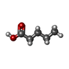  Mass: 102.132 Da / Num. of mol.: 2 / Source method: obtained synthetically / Formula: C5H10O2 Mass: 102.132 Da / Num. of mol.: 2 / Source method: obtained synthetically / Formula: C5H10O2#3: Water | ChemComp-HOH / |  Mass: 18.015 Da / Num. of mol.: 125 / Source method: isolated from a natural source / Formula: H2O Mass: 18.015 Da / Num. of mol.: 125 / Source method: isolated from a natural source / Formula: H2OHas ligand of interest | N | Has protein modification | N | |
|---|
-Experimental details
-Experiment
| Experiment | Method: X-RAY DIFFRACTION / Number of used crystals: 1 |
|---|
- Sample preparation
| Crystal | Density Matthews: 2.03 Å3/Da / Density % sol: 39.35 % |
|---|---|
| Crystal grow | Temperature: 293 K / Method: vapor diffusion / pH: 8.5 Details: 0.1 M Tris (pH 8.5), 0.1 M Mg formate, and 30% PE14/4, with 10% glycerol used as cryoprotectant |
-Data collection
| Diffraction | Mean temperature: 100 K / Serial crystal experiment: N |
|---|---|
| Diffraction source | Source: SYNCHROTRON / Site: Australian Synchrotron / Beamline: MX2 / Wavelength: 0.95373 Å |
| Detector | Type: DECTRIS EIGER X 16M / Detector: PIXEL / Date: Dec 1, 2025 |
| Radiation | Protocol: SINGLE WAVELENGTH / Monochromatic (M) / Laue (L): M / Scattering type: x-ray |
| Radiation wavelength | Wavelength: 0.95373 Å / Relative weight: 1 |
| Reflection | Resolution: 2.12→47.7 Å / Num. obs: 24828 / % possible obs: 98.6 % / Redundancy: 3.8 % / Biso Wilson estimate: 34.45 Å2 / CC1/2: 0.999 / Rpim(I) all: 0.026 / Net I/σ(I): 12.7 |
| Reflection shell | Resolution: 2.12→2.18 Å / Mean I/σ(I) obs: 3.7 / Num. unique obs: 1949 / CC1/2: 0.95 / Rpim(I) all: 0.152 |
- Processing
| Software |
| |||||||||||||||||||||||||||||||||||||||||||||||||||||||||||||||||||||||||||||||||||||||||||||||||||||||||
|---|---|---|---|---|---|---|---|---|---|---|---|---|---|---|---|---|---|---|---|---|---|---|---|---|---|---|---|---|---|---|---|---|---|---|---|---|---|---|---|---|---|---|---|---|---|---|---|---|---|---|---|---|---|---|---|---|---|---|---|---|---|---|---|---|---|---|---|---|---|---|---|---|---|---|---|---|---|---|---|---|---|---|---|---|---|---|---|---|---|---|---|---|---|---|---|---|---|---|---|---|---|---|---|---|---|---|
| Refinement | Method to determine structure: MOLECULAR REPLACEMENT / Resolution: 2.12→40.76 Å / SU ML: 0.1937 / Cross valid method: FREE R-VALUE / σ(F): 0.03 / Phase error: 24.4591 Stereochemistry target values: GeoStd + Monomer Library + CDL v1.2
| |||||||||||||||||||||||||||||||||||||||||||||||||||||||||||||||||||||||||||||||||||||||||||||||||||||||||
| Solvent computation | Shrinkage radii: 0.9 Å / VDW probe radii: 1.1 Å / Solvent model: FLAT BULK SOLVENT MODEL | |||||||||||||||||||||||||||||||||||||||||||||||||||||||||||||||||||||||||||||||||||||||||||||||||||||||||
| Displacement parameters | Biso mean: 47.96 Å2 | |||||||||||||||||||||||||||||||||||||||||||||||||||||||||||||||||||||||||||||||||||||||||||||||||||||||||
| Refinement step | Cycle: LAST / Resolution: 2.12→40.76 Å
| |||||||||||||||||||||||||||||||||||||||||||||||||||||||||||||||||||||||||||||||||||||||||||||||||||||||||
| Refine LS restraints |
| |||||||||||||||||||||||||||||||||||||||||||||||||||||||||||||||||||||||||||||||||||||||||||||||||||||||||
| LS refinement shell |
|
