 Movie
Movie Controller

[English] 日本語
 Yorodumi
- PDB-9zj8: Crystal structure of MERS 3CL protease in complex with inhibitor ... -
+ Open data

- Basic information
| Entry | Database: PDB / ID: 9zj8 | |||||||||
|---|---|---|---|---|---|---|---|---|---|---|
| Title | Crystal structure of MERS 3CL protease in complex with inhibitor AMJ-II-72 | |||||||||
 Components Components | 3C-like proteinase nsp5 | |||||||||
 Keywords Keywords | HYDROLASE/HYDROLASE INHIBITOR / HYDROLASE / HYDROLASE-HYDROLASE INHIBITOR complex | |||||||||
| Function / homology |  Function and homology information Function and homology informationhost cell membrane / Hydrolases; Glycosylases; Hydrolysing N-glycosyl compounds / viral genome replication / methyltransferase activity / endonuclease activity / methylation / SARS coronavirus main proteinase / symbiont-mediated degradation of host mRNA / mRNA guanylyltransferase / symbiont-mediated suppression of host ISG15-protein conjugation ...host cell membrane / Hydrolases; Glycosylases; Hydrolysing N-glycosyl compounds / viral genome replication / methyltransferase activity / endonuclease activity / methylation / SARS coronavirus main proteinase / symbiont-mediated degradation of host mRNA / mRNA guanylyltransferase / symbiont-mediated suppression of host ISG15-protein conjugation / G-quadruplex RNA binding / mRNA guanylyltransferase activity / symbiont-mediated suppression of host cytoplasmic pattern recognition receptor signaling pathway via inhibition of IRF3 activity / omega peptidase activity / symbiont-mediated perturbation of host ubiquitin-like protein modification / ubiquitinyl hydrolase 1 / Hydrolases; Acting on peptide bonds (peptidases); Cysteine endopeptidases / cysteine-type deubiquitinase activity / single-stranded RNA binding / viral protein processing / host cell perinuclear region of cytoplasm / symbiont-mediated suppression of host type I interferon-mediated signaling pathway / symbiont-mediated suppression of host gene expression / viral translational frameshifting / symbiont-mediated activation of host autophagy / cysteine-type endopeptidase activity / proteolysis / zinc ion binding Similarity search - Function | |||||||||
| Biological species |  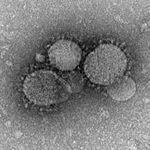 Middle East respiratory syndrome-related coronavirus Middle East respiratory syndrome-related coronavirus | |||||||||
| Method |  X-RAY DIFFRACTION / SYNCHROTRON / MOLECULAR REPLACEMENT / Resolution: 2.1 Å X-RAY DIFFRACTION / SYNCHROTRON / MOLECULAR REPLACEMENT / Resolution: 2.1 Å | |||||||||
 Authors Authors | Liu, L. / Lovell, S. / Battaile, K.P. / Jesri, A.R.M. / Groutas, W.C. | |||||||||
| Funding support |  United States, 2items United States, 2items
| |||||||||
 Citation Citation | Journal: Eur.J.Med.Chem. / Year: 2026 Title: Structure-guided design of broad-spectrum inhibitors of coronaviral proteases embodying a 1,3,2-oxazaphospholidin-3-one scaffold as a versatile design element. Authors: Nguyen, H.N. / Ranasinghe, P.S. / Ilesinghe, I.K.R.S. / Dampalla, C.S. / Rathnayake, A.D. / Liska, Z. / Jesri, A.M. / Azmi, Z. / Nevonen, D.E. / Kim, Y. / Ung, A.R. / Taylor, K.E. / Cooper, ...Authors: Nguyen, H.N. / Ranasinghe, P.S. / Ilesinghe, I.K.R.S. / Dampalla, C.S. / Rathnayake, A.D. / Liska, Z. / Jesri, A.M. / Azmi, Z. / Nevonen, D.E. / Kim, Y. / Ung, A.R. / Taylor, K.E. / Cooper, A. / Liu, L. / Battaile, K.P. / Thurman, H.A. / Gusachenko, E. / Lovell, S. / Groutas, W.C. / Chang, K.O. | |||||||||
| History |
|
- Structure visualization
| Structure viewer | Molecule: MolmilJmol/JSmol |
|---|
- Downloads & links
-Download
| PDBx/mmCIF format | 9zj8.cif.gz | 131.1 KB | Display | PDBx/mmCIF format |
|---|---|---|---|---|
| PDB format | pdb9zj8.ent.gz | Display | PDB format | |
| PDBx/mmJSON format | 9zj8.json.gz | Tree view | PDBx/mmJSON format | |
| Others |  Other downloads Other downloads |
-Validation report
| Arichive directory | https://data.pdbj.org/pub/pdb/validation_reports/zj/9zj8ftp://data.pdbj.org/pub/pdb/validation_reports/zj/9zj8 | HTTPS FTP |
|---|
-Related structure data
| Related structure data |  9zj1C  9zj2C  9zj3C  9zj4C  9zj5C  9zj6C  9zjbC  9zjcC  9zzhC C: citing same article ( |
|---|---|
| Similar structure data |
-Links
 PDBj
PDBj
- Assembly
| Deposited unit | 
| ||||||||
|---|---|---|---|---|---|---|---|---|---|
| 1 | 
| ||||||||
| Unit cell |
|
-Components
| #1: Protein | Mass: 34314.242 Da / Num. of mol.: 1 / Fragment: UNP residues 3248-3553 Source method: isolated from a genetically manipulated source Source: (gene. exp.) Middle East respiratory syndrome-related coronavirusGene: 1a / Plasmid: pET-28 / Production host:   Escherichia coli BL21(DE3) (bacteria) Escherichia coli BL21(DE3) (bacteria)References: UniProt: K9N638, SARS coronavirus main proteinase |
|---|---|
| #2: Chemical | ChemComp-A1C2N / Mass: 612.627 Da / Num. of mol.: 1 / Source method: obtained synthetically / Formula: C28H42FN4O8P / Feature type: SUBJECT OF INVESTIGATION |
| #3: Water | ChemComp-HOH /  Mass: 18.015 Da / Num. of mol.: 48 / Source method: isolated from a natural source / Formula: H2O Mass: 18.015 Da / Num. of mol.: 48 / Source method: isolated from a natural source / Formula: H2O |
| Has ligand of interest | Y |
| Has protein modification | Y |
-Experimental details
-Experiment
| Experiment | Method: X-RAY DIFFRACTION / Number of used crystals: 1 |
|---|
- Sample preparation
| Crystal | Density Matthews: 1.94 Å3/Da / Density % sol: 36.51 % |
|---|---|
| Crystal grow | Temperature: 291 K / Method: vapor diffusion, sitting drop / pH: 6.5 Details: 25% (w/v) PEG 3350, 100 mM Bis-Tris pH 6.5, 200 mM Magnesium Chloride |
-Data collection
| Diffraction | Mean temperature: 100 K / Serial crystal experiment: N |
|---|---|
| Diffraction source | Source: SYNCHROTRON / Site: NSLS-II / Beamline: 19-ID / Wavelength: 0.97956 Å |
| Detector | Type: DECTRIS EIGER2 XE 9M / Detector: PIXEL / Date: Mar 24, 2022 |
| Radiation | Monochromator: Double Crystal Si 111 / Protocol: SINGLE WAVELENGTH / Monochromatic (M) / Laue (L): M / Scattering type: x-ray |
| Radiation wavelength | Wavelength: 0.97956 Å / Relative weight: 1 |
| Reflection | Resolution: 2.1→49.14 Å / Num. obs: 14719 / % possible obs: 95.4 % / Redundancy: 3.5 % / CC1/2: 0.997 / Rmerge(I) obs: 0.09 / Rpim(I) all: 0.056 / Rrim(I) all: 0.106 / Χ2: 0.96 / Net I/σ(I): 9.7 / Num. measured all: 51391 |
| Reflection shell | Resolution: 2.1→2.16 Å / % possible obs: 99.5 % / Redundancy: 3.7 % / Rmerge(I) obs: 0.654 / Num. measured all: 4636 / Num. unique obs: 1254 / CC1/2: 0.697 / Rpim(I) all: 0.393 / Rrim(I) all: 0.765 / Χ2: 0.89 / Net I/σ(I) obs: 2 |
- Processing
| Software |
| ||||||||||||||||||||||||||||||||||||||||||||||||||||||||||||||||||||||||||||||||||||||||||||||||||||
|---|---|---|---|---|---|---|---|---|---|---|---|---|---|---|---|---|---|---|---|---|---|---|---|---|---|---|---|---|---|---|---|---|---|---|---|---|---|---|---|---|---|---|---|---|---|---|---|---|---|---|---|---|---|---|---|---|---|---|---|---|---|---|---|---|---|---|---|---|---|---|---|---|---|---|---|---|---|---|---|---|---|---|---|---|---|---|---|---|---|---|---|---|---|---|---|---|---|---|---|---|---|
| Refinement | Method to determine structure: MOLECULAR REPLACEMENT / Resolution: 2.1→46.37 Å / SU ML: 0.27 / Cross valid method: FREE R-VALUE / σ(F): 1.05 / Phase error: 27.89 / Stereochemistry target values: ML
| ||||||||||||||||||||||||||||||||||||||||||||||||||||||||||||||||||||||||||||||||||||||||||||||||||||
| Solvent computation | Shrinkage radii: 0.9 Å / VDW probe radii: 1.11 Å / Solvent model: FLAT BULK SOLVENT MODEL | ||||||||||||||||||||||||||||||||||||||||||||||||||||||||||||||||||||||||||||||||||||||||||||||||||||
| Refinement step | Cycle: LAST / Resolution: 2.1→46.37 Å
| ||||||||||||||||||||||||||||||||||||||||||||||||||||||||||||||||||||||||||||||||||||||||||||||||||||
| Refine LS restraints |
| ||||||||||||||||||||||||||||||||||||||||||||||||||||||||||||||||||||||||||||||||||||||||||||||||||||
| LS refinement shell |
| ||||||||||||||||||||||||||||||||||||||||||||||||||||||||||||||||||||||||||||||||||||||||||||||||||||
| Refinement TLS params. | Method: refined / Refine-ID: X-RAY DIFFRACTION
| ||||||||||||||||||||||||||||||||||||||||||||||||||||||||||||||||||||||||||||||||||||||||||||||||||||
| Refinement TLS group |
|
