National Institutes of Health/National Institute Of Allergy and Infectious Diseases (NIH/NIAID)
AI144462
United States
Bill & Melinda Gates Foundation
INV-007522
United States
Bill & Melinda Gates Foundation
INV-008813
United States
Citation
Journal: Proc Natl Acad Sci U S A / Year: 2026 Title: Structural basis of membrane engagement and polyreactivity control in HIV-1 MPER broadly neutralizing antibodies. Authors: So Yeon Cho / Kimmo Rantalainen / Gabriel Ozorowski / Danny Lu / Ryan Tingle / Wen-Hsin Lee / Andrew B Ward / William R Schief / Ian A Wilson / Abstract: The membrane-proximal external region (MPER) of HIV-1 Env represents a critical target for broadly neutralizing antibodies (bnAbs) due to its conservation and functional importance. However, MPER- ...The membrane-proximal external region (MPER) of HIV-1 Env represents a critical target for broadly neutralizing antibodies (bnAbs) due to its conservation and functional importance. However, MPER-targeting bnAbs recognize composite epitopes comprising peptide and viral membrane lipid components, creating an inherent tension between viral neutralization efficacy and polyreactivity. 10E8-class antibodies exhibit high neutralization potency with low polyreactivity, whereas 4E10-class antibodies show comparably broad neutralization but higher polyreactivity, underscoring the need to understand the structural basis of this distinction. We therefore determined crystal structures of DH511.1 (memory B cell-derived), DH511.12P (plasma cell-derived), and VRC42.01 in complex with MPER peptide and phosphatidic acid, along with a cryo-EM reconstruction of DH511.2 bound to membrane-embedded Env. Through integrative analysis taking into account previously determined structures of other MPER bnAbs, we reveal two distinct lipid recognition strategies. Groove-mediated binders, including 10E8 and DH511, engage lipids through antibody-membrane interface grooves with distinct geometries and angular approaches to the membrane. In contrast, heavy chain-mediated binders, including 4E10, PGZL1, and VRC42, utilize positively charged CDR H1 patches for direct lipid headgroup recognition. Importantly, DH511 lineage members exhibited differential cardiolipin polyreactivity linked to their maturation stage. PGZL1 and VRC42.01 employ weaker positive patches at lipid-binding sites than 4E10, and PGZL1 additionally introduces a CDR H3-mediated negative patch that creates electrostatic repulsion with negatively charged lipid headgroups, thereby limiting nonspecific interactions. These findings provide a structural framework for understanding how MPER bnAbs balance lipid binding with specificity and inform immunogen design for inducing safe and effective neutralizing responses.
Mass: 24051.127 Da / Num. of mol.: 1 Source method: isolated from a genetically manipulated source Source: (gene. exp.) Homo sapiens (human) / Production host: Homo sapiens (human)
#2: Antibody
AntibodyVRC42.01Fablightchain
Mass: 23249.666 Da / Num. of mol.: 1 Source method: isolated from a genetically manipulated source Source: (gene. exp.) Homo sapiens (human) / Production host: Homo sapiens (human)
In the structure databanks used in Yorodumi, some data are registered as the other names, "COVID-19 virus" and "2019-nCoV". Here are the details of the virus and the list of structure data.
Jan 31, 2019. EMDB accession codes are about to change! (news from PDBe EMDB page)
EMDB accession codes are about to change! (news from PDBe EMDB page)
The allocation of 4 digits for EMDB accession codes will soon come to an end. Whilst these codes will remain in use, new EMDB accession codes will include an additional digit and will expand incrementally as the available range of codes is exhausted. The current 4-digit format prefixed with “EMD-” (i.e. EMD-XXXX) will advance to a 5-digit format (i.e. EMD-XXXXX), and so on. It is currently estimated that the 4-digit codes will be depleted around Spring 2019, at which point the 5-digit format will come into force.
The EM Navigator/Yorodumi systems omit the EMD- prefix.
Related info.:Q: What is EMD? / ID/Accession-code notation in Yorodumi/EM Navigator
Yorodumi is a browser for structure data from EMDB, PDB, SASBDB, etc.
This page is also the successor to EM Navigator detail page, and also detail information page/front-end page for Omokage search.
The word "yorodu" (or yorozu) is an old Japanese word meaning "ten thousand". "mi" (miru) is to see.
Related info.:EMDB / PDB / SASBDB / Comparison of 3 databanks / Yorodumi Search / Aug 31, 2016. New EM Navigator & Yorodumi / Yorodumi Papers / Jmol/JSmol / Function and homology information / Changes in new EM Navigator and Yorodumi
 Movie
Movie Controller

 Yorodumi
 Open data
 Basic information
 Components
 Keywords
 Function and homology information
 Homo sapiens (human)
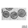 Human immunodeficiency virus 1
 X-RAY DIFFRACTION /
  Authors
 United States, 3items
  Citation
 Structure visualization
 Downloads & links
 Other downloads


 PDBj
PDBj

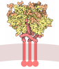
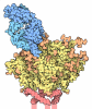
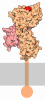
 Assembly
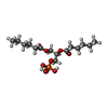
 Mass: 368.360 Da / Num. of mol.: 2 / Source method: obtained synthetically / Formula: C15H29O8P / Feature type: SUBJECT OF INVESTIGATION / Comment: phospholipid*YM
 Sample preparation
 Processing