 Movie
Movie Controller

[English] 日本語
 Yorodumi
- PDB-9zei: The 200-K crystal structure of CYP199A4 bound to 4-phenoxybenzoic... -
+ Open data

- Basic information
| Entry | Database: PDB / ID: 9zei | ||||||
|---|---|---|---|---|---|---|---|
| Title | The 200-K crystal structure of CYP199A4 bound to 4-phenoxybenzoic acid (dataset 3; increasing temperature series) | ||||||
 Components Components | Cytochrome P450 | ||||||
 Keywords Keywords | OXIDOREDUCTASE / Cytochrome P450 / variable-temperature crystallography | ||||||
| Function / homology |  Function and homology information Function and homology informationoxidoreductase activity, acting on paired donors, with incorporation or reduction of molecular oxygen / monooxygenase activity / iron ion binding / heme binding Similarity search - Function | ||||||
| Biological species |  Rhodopseudomonas palustris HaA2 (phototrophic) Rhodopseudomonas palustris HaA2 (phototrophic) | ||||||
| Method |  X-RAY DIFFRACTION / SYNCHROTRON / MOLECULAR REPLACEMENT / Resolution: 1.74 Å X-RAY DIFFRACTION / SYNCHROTRON / MOLECULAR REPLACEMENT / Resolution: 1.74 Å | ||||||
 Authors Authors | Podgorski, M.N. / Bell, S.G. | ||||||
| Funding support |  Australia, 1items Australia, 1items
| ||||||
 Citation Citation | Journal: Chem Sci / Year: 2026 Title: Structural basis for substrate-dependent allostery in oxygen activation by a cytochrome P450 enzyme revealed by analysis at different temperatures. Authors: Podgorski, M.N. / McDougal, D.P. / Campbell, E.C. / Bruning, J.B. / Bell, S.G. | ||||||
| History |
|
- Structure visualization
| Structure viewer | Molecule: MolmilJmol/JSmol |
|---|
- Downloads & links
-Download
| PDBx/mmCIF format | 9zei.cif.gz | 103.9 KB | Display | PDBx/mmCIF format |
|---|---|---|---|---|
| PDB format | pdb9zei.ent.gz | 75.5 KB | Display | PDB format |
| PDBx/mmJSON format | 9zei.json.gz | Tree view | PDBx/mmJSON format | |
| Others |  Other downloads Other downloads |
-Validation report
| Arichive directory | https://data.pdbj.org/pub/pdb/validation_reports/ze/9zeiftp://data.pdbj.org/pub/pdb/validation_reports/ze/9zei | HTTPS FTP |
|---|
-Related structure data
| Related structure data |  9doeC  9mimC  9mioC  9mjeC  9mjfC  9mjjC  9mjkC  9plsC  9pmaC  9pmcC  9zegC  9zehC C: citing same article ( |
|---|---|
| Similar structure data |
-Links
 PDBj
PDBj
- Assembly
| Deposited unit | 
| ||||||||
|---|---|---|---|---|---|---|---|---|---|
| 1 |
| ||||||||
| Unit cell |
|
-Components
| #1: Protein | Mass: 44587.430 Da / Num. of mol.: 1 Source method: isolated from a genetically manipulated source Source: (gene. exp.) Rhodopseudomonas palustris HaA2 (phototrophic)Gene: RPB_3613 / Production host: |
|---|---|
| #2: Chemical | ChemComp-JO4 / 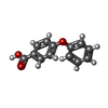  Mass: 214.217 Da / Num. of mol.: 1 / Source method: obtained synthetically / Formula: C13H10O3 / Feature type: SUBJECT OF INVESTIGATION Mass: 214.217 Da / Num. of mol.: 1 / Source method: obtained synthetically / Formula: C13H10O3 / Feature type: SUBJECT OF INVESTIGATION |
| #3: Chemical | ChemComp-HEM /   Mass: 616.487 Da / Num. of mol.: 1 / Source method: obtained synthetically / Formula: C34H32FeN4O4 Mass: 616.487 Da / Num. of mol.: 1 / Source method: obtained synthetically / Formula: C34H32FeN4O4 |
| #4: Chemical | ChemComp-CL /   Mass: 35.453 Da / Num. of mol.: 1 / Source method: obtained synthetically / Formula: Cl Mass: 35.453 Da / Num. of mol.: 1 / Source method: obtained synthetically / Formula: Cl |
| #5: Water | ChemComp-HOH /  Mass: 18.015 Da / Num. of mol.: 378 / Source method: isolated from a natural source / Formula: H2O Mass: 18.015 Da / Num. of mol.: 378 / Source method: isolated from a natural source / Formula: H2O |
| Has ligand of interest | Y |
| Has protein modification | N |
-Experimental details
-Experiment
| Experiment | Method: X-RAY DIFFRACTION / Number of used crystals: 1 |
|---|
- Sample preparation
| Crystal | Density Matthews: 2.04 Å3/Da / Density % sol: 39.73 % |
|---|---|
| Crystal grow | Temperature: 289.15 K / Method: vapor diffusion, hanging drop / pH: 7.4 Details: The crystallization buffer was 100 mM Bis-Tris buffer (adjusted to pH 5.0-5.75 with acetic acid), 0.2 M magnesium acetate and 20-32% PEG 3350 PH range: 5.0-5.75 |
-Data collection
| Diffraction | Mean temperature: 200 K / Serial crystal experiment: N |
|---|---|
| Diffraction source | Source: SYNCHROTRON / Site: Australian Synchrotron / Beamline: MX2 / Wavelength: 0.95373 Å |
| Detector | Type: DECTRIS EIGER X 16M / Detector: PIXEL / Date: Oct 6, 2021 |
| Radiation | Monochromator: Double-crystal Si(111) liquid nitrogen cooled (DC) or channel-cut Si(111) liquid nitrogen cooled (CC) Protocol: SINGLE WAVELENGTH / Monochromatic (M) / Laue (L): M / Scattering type: x-ray |
| Radiation wavelength | Wavelength: 0.95373 Å / Relative weight: 1 |
| Reflection | Resolution: 1.74→44.42 Å / Num. obs: 37236 / % possible obs: 99.5 % / Redundancy: 6.8 % / CC1/2: 0.998 / Rmerge(I) obs: 0.107 / Rpim(I) all: 0.044 / Rrim(I) all: 0.115 / Χ2: 0.5 / Net I/σ(I): 9.3 / Num. measured all: 253410 |
| Reflection shell | Resolution: 1.74→1.77 Å / % possible obs: 96.3 % / Redundancy: 6.6 % / Rmerge(I) obs: 1.398 / Num. measured all: 12825 / Num. unique obs: 1944 / CC1/2: 0.608 / Rpim(I) all: 0.582 / Rrim(I) all: 1.518 / Χ2: 0.4 / Net I/σ(I) obs: 1 |
- Processing
| Software |
| ||||||||||||||||||||||||||||||||||||||||||||||||||||||||||||||||||||||||||||||||||||||||||||||||||
|---|---|---|---|---|---|---|---|---|---|---|---|---|---|---|---|---|---|---|---|---|---|---|---|---|---|---|---|---|---|---|---|---|---|---|---|---|---|---|---|---|---|---|---|---|---|---|---|---|---|---|---|---|---|---|---|---|---|---|---|---|---|---|---|---|---|---|---|---|---|---|---|---|---|---|---|---|---|---|---|---|---|---|---|---|---|---|---|---|---|---|---|---|---|---|---|---|---|---|---|
| Refinement | Method to determine structure: MOLECULAR REPLACEMENT / Resolution: 1.74→43.29 Å / SU ML: 0.19 / Cross valid method: FREE R-VALUE / σ(F): 1.36 / Phase error: 20.87 / Stereochemistry target values: ML
| ||||||||||||||||||||||||||||||||||||||||||||||||||||||||||||||||||||||||||||||||||||||||||||||||||
| Solvent computation | Shrinkage radii: 0.9 Å / VDW probe radii: 1.1 Å / Solvent model: FLAT BULK SOLVENT MODEL | ||||||||||||||||||||||||||||||||||||||||||||||||||||||||||||||||||||||||||||||||||||||||||||||||||
| Refinement step | Cycle: LAST / Resolution: 1.74→43.29 Å
| ||||||||||||||||||||||||||||||||||||||||||||||||||||||||||||||||||||||||||||||||||||||||||||||||||
| Refine LS restraints |
| ||||||||||||||||||||||||||||||||||||||||||||||||||||||||||||||||||||||||||||||||||||||||||||||||||
| LS refinement shell |
|
