 Movie
Movie Controller

[English] 日本語
 Yorodumi
- PDB-9zbt: Visualization of PriA/PriB/DnaT complexes reveals mechanisms gove... -
+ Open data

- Basic information
| Entry | Database: PDB / ID: 9zbt | ||||||||||||||||||||||||
|---|---|---|---|---|---|---|---|---|---|---|---|---|---|---|---|---|---|---|---|---|---|---|---|---|---|
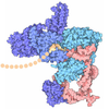
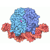
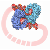
| Title | Visualization of PriA/PriB/DnaT complexes reveals mechanisms governing structure-specific assembly of the DNA replication restart primosome | ||||||||||||||||||||||||
 Components Components |
| ||||||||||||||||||||||||
 Keywords Keywords | REPLICATION / DNA replication / helicase / oligomer / DNA repair | ||||||||||||||||||||||||
| Function / homology |  Function and homology information Function and homology informationpre-primosome complex / DnaB-DnaC-DnaT-PriA-PriC complex / DnaB-DnaC-DnaT-PriA-PriB complex / plasmid maintenance / primosome complex / DNA replication, synthesis of primer / protein homotrimerization / DNA 3'-5' helicase / DNA replication initiation / 3'-5' DNA helicase activity ...pre-primosome complex / DnaB-DnaC-DnaT-PriA-PriC complex / DnaB-DnaC-DnaT-PriA-PriB complex / plasmid maintenance / primosome complex / DNA replication, synthesis of primer / protein homotrimerization / DNA 3'-5' helicase / DNA replication initiation / 3'-5' DNA helicase activity / helicase activity / response to gamma radiation / replication fork processing / response to radiation / DNA-templated DNA replication / double-strand break repair / single-stranded DNA binding / DNA recombination / DNA replication / response to antibiotic / magnesium ion binding / ATP hydrolysis activity / DNA binding / zinc ion binding / ATP binding / identical protein binding Similarity search - Function | ||||||||||||||||||||||||
| Biological species |   Escherichia coli K-12 (bacteria) Escherichia coli K-12 (bacteria)synthetic construct (others) | ||||||||||||||||||||||||
| Method | ELECTRON MICROSCOPY / single particle reconstruction / cryo EM / Resolution: 3.22 Å | ||||||||||||||||||||||||
 Authors Authors | Duckworth, A.T. / Keck, J.L. / Grant, T. | ||||||||||||||||||||||||
| Funding support |  United States, 1items United States, 1items
| ||||||||||||||||||||||||
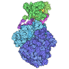
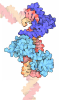
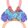
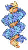
 Citation Citation | Journal: Nat Commun / Year: 2026 Title: Visualization of the complete preprimosome reveals the structural mechanisms governing DNA replication restart. Authors: Peter L Ducos / Alexander T Duckworth / Kenneth A Satyshur / James L Keck / Timothy Grant / Abstract: Replication restart pathways reinitiate DNA replication processes following their premature termination. In Escherichia coli, this essential process begins with regulated assembly of the preprimosome ...Replication restart pathways reinitiate DNA replication processes following their premature termination. In Escherichia coli, this essential process begins with regulated assembly of the preprimosome complex, comprising the PriA, PriB, and DnaT proteins, onto an abandoned replication fork. Here, we present two distinct preprimosome structures. One represents an intermediate stage in preprimosome assembly with a single DnaT C-terminal domain (DnaT) bound to PriA/PriB/DNA. The second captures the mature preprimosome, in which filamentation of multiple DnaT molecules catalyzes the handoff of the single-stranded lagging-strand DNA from PriB to DnaT. The DnaT N-terminal domain forms a separate, independent oligomer in the mature structure. Taken together, our results detail the molecular mechanisms underlying replication restart initiation and regulation and suggest mechanistic similarities between DnaT and the canonical initiator protein DnaA. | ||||||||||||||||||||||||
| History |
|
- Structure visualization
| Structure viewer | Molecule: MolmilJmol/JSmol |
|---|
- Downloads & links
-Download
| PDBx/mmCIF format | 9zbt.cif.gz | 228.8 KB | Display | PDBx/mmCIF format |
|---|---|---|---|---|
| PDB format | pdb9zbt.ent.gz | Display | PDB format | |
| PDBx/mmJSON format | 9zbt.json.gz | Tree view | PDBx/mmJSON format | |
| Others |  Other downloads Other downloads |
-Validation report
| Arichive directory | https://data.pdbj.org/pub/pdb/validation_reports/zb/9zbtftp://data.pdbj.org/pub/pdb/validation_reports/zb/9zbt | HTTPS FTP |
|---|
-Related structure data
| Related structure data |  74008MC  9zbuC M: map data used to model this data C: citing same article ( |
|---|---|
| Similar structure data |

-Links
 PDBj
PDBj




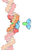


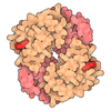
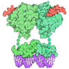



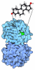

- Assembly
| Deposited unit | 
|
|---|---|
| 1 |
|
-Components
-Replication restart protein ... , 2 types, 3 molecules DAB
| #1: Protein | Mass: 19474.025 Da / Num. of mol.: 1 Source method: isolated from a genetically manipulated source Source: (gene. exp.) Escherichia coli K-12 (bacteria) / Gene: dnaT, b4362, JW4326 / Production host: Escherichia coli BL21(DE3) (bacteria) / References: UniProt: P0A8J2 |
|---|---|
| #2: Protein | Mass: 11459.194 Da / Num. of mol.: 2 Source method: isolated from a genetically manipulated source Source: (gene. exp.) Escherichia coli K-12 (bacteria) / Gene: priB, b4201, JW4159 / Production host: Escherichia coli BL21(DE3) (bacteria) / References: UniProt: P07013 |
-DNA chain , 3 types, 3 molecules GJZ
| #3: DNA chain | Mass: 12206.812 Da / Num. of mol.: 1 / Source method: obtained synthetically / Source: (synth.) synthetic construct (others) |
|---|---|
| #5: DNA chain | Mass: 12399.983 Da / Num. of mol.: 1 / Source method: obtained synthetically / Source: (synth.) synthetic construct (others) |
| #6: DNA chain | Mass: 4567.944 Da / Num. of mol.: 1 / Source method: obtained synthetically / Source: (synth.) synthetic construct (others) |
-Protein / Non-polymers , 2 types, 3 molecules H
| #4: Protein | Mass: 81765.820 Da / Num. of mol.: 1 Source method: isolated from a genetically manipulated source Source: (gene. exp.) Escherichia coli K-12 (bacteria) / Gene: priA, b3935, JW3906 / Production host: Escherichia coli BL21(DE3) (bacteria)References: UniProt: P17888, Hydrolases; Acting on acid anhydrides; Acting on acid anhydrides to facilitate cellular and subcellular movement |
|---|---|
| #7: Chemical |  Mass: 65.409 Da / Num. of mol.: 2 / Source method: obtained synthetically / Formula: Zn Mass: 65.409 Da / Num. of mol.: 2 / Source method: obtained synthetically / Formula: Zn |
-Details
| Has ligand of interest | N |
|---|---|
| Has protein modification | N |
-Experimental details
-Experiment
| Experiment | Method: ELECTRON MICROSCOPY |
|---|---|
| EM experiment | Aggregation state: PARTICLE / 3D reconstruction method: single particle reconstruction |
- Sample preparation
| Component | Name: Complex of PriA, PriB, DnaT, and replication fork / Type: COMPLEX / Entity ID: #2, #1, #4, #3, #5-#6 / Source: RECOMBINANT |
|---|---|
| Molecular weight | Experimental value: NO |
| Source (natural) | Organism: Escherichia coli K-12 (bacteria) |
| Source (recombinant) | Organism: Escherichia coli BL21(DE3) (bacteria) |
| Buffer solution | pH: 8 Details: 50 mM Tris-HCl, pH 8, 2 mM dithiothreitol, 5 mM ethylenediaminetetraacetic acid, 75 mM NaCl |
| Specimen | Conc.: 1 mg/ml / Embedding applied: NO / Shadowing applied: NO / Staining applied: NO / Vitrification applied: YES |
| Specimen support | Grid material: GOLD / Grid mesh size: 300 divisions/in. / Grid type: UltrAuFoil R1.2/1.3 |
| Vitrification | Instrument: FEI VITROBOT MARK IV / Cryogen name: ETHANE / Humidity: 100 % / Chamber temperature: 277 K |
- Electron microscopy imaging
| Experimental equipment |  Model: Titan Krios / Image courtesy: FEI Company | |||||||||||||||||||||
|---|---|---|---|---|---|---|---|---|---|---|---|---|---|---|---|---|---|---|---|---|---|---|
| Microscopy | Model: TFS KRIOS | |||||||||||||||||||||
| Electron gun | Electron source:  FIELD EMISSION GUN / Accelerating voltage: 300 kV / Illumination mode: FLOOD BEAM FIELD EMISSION GUN / Accelerating voltage: 300 kV / Illumination mode: FLOOD BEAM | |||||||||||||||||||||
| Electron lens | Mode: BRIGHT FIELD / Nominal defocus max: 2500 nm / Nominal defocus min: 500 nm | |||||||||||||||||||||
| Specimen holder | Cryogen: NITROGEN | |||||||||||||||||||||
| Image recording |
| |||||||||||||||||||||
| EM imaging optics | Energyfilter name: GIF Bioquantum / Energyfilter slit width: 20 eV |
- Processing
| EM software |
| |||||||||||||||||||||||||||||||||
|---|---|---|---|---|---|---|---|---|---|---|---|---|---|---|---|---|---|---|---|---|---|---|---|---|---|---|---|---|---|---|---|---|---|---|
| CTF correction | Type: PHASE FLIPPING AND AMPLITUDE CORRECTION | |||||||||||||||||||||||||||||||||
| Particle selection | Num. of particles selected: 2200000 | |||||||||||||||||||||||||||||||||
| 3D reconstruction | Resolution: 3.22 Å / Resolution method: FSC 0.143 CUT-OFF / Num. of particles: 857000 / Num. of class averages: 15 / Symmetry type: POINT | |||||||||||||||||||||||||||||||||
| Refinement | Highest resolution: 3.22 Å Stereochemistry target values: REAL-SPACE (WEIGHTED MAP SUM AT ATOM CENTERS) | |||||||||||||||||||||||||||||||||
| Refine LS restraints |
|
