 Movie
Movie Controller

[English] 日本語
 Yorodumi
- PDB-9zag: Crystal structure of a glyceraldehyde-3-phosphate dehydrogenase f... -
+ Open data

- Basic information
| Entry | Database: PDB / ID: 9zag | |||||||||
|---|---|---|---|---|---|---|---|---|---|---|
| Title | Crystal structure of a glyceraldehyde-3-phosphate dehydrogenase from Neisseria gonorrhoeae in complex with NAD and GLYCERALDEHYDE-3-PHOSPHATE | |||||||||
 Components Components | Glyceraldehyde-3-phosphate dehydrogenase | |||||||||
 Keywords Keywords | OXIDOREDUCTASE / SSGCID / STRUCTURAL GENOMICS / SEATTLE STRUCTURAL GENOMICS CENTER FOR INFECTIOUS DISEASE / glyceraldehyde-3-phosphate dehydrogenase | |||||||||
| Function / homology |  Function and homology information Function and homology informationOxidoreductases; Acting on the aldehyde or oxo group of donors; With NAD+ or NADP+ as acceptor / oxidoreductase activity, acting on the aldehyde or oxo group of donors, NAD or NADP as acceptor / glucose metabolic process / NAD binding / NADP binding Similarity search - Function | |||||||||
| Biological species |  Neisseria gonorrhoeae NCCP11945 (bacteria) Neisseria gonorrhoeae NCCP11945 (bacteria) | |||||||||
| Method |  X-RAY DIFFRACTION / SYNCHROTRON / MOLECULAR REPLACEMENT / Resolution: 1.91 Å X-RAY DIFFRACTION / SYNCHROTRON / MOLECULAR REPLACEMENT / Resolution: 1.91 Å | |||||||||
 Authors Authors | Seattle Structural Genomics Center for Infectious Disease / Seattle Structural Genomics Center for Infectious Disease (SSGCID) | |||||||||
| Funding support |  United States, 2items United States, 2items
| |||||||||
 Citation Citation | Journal: To be published Title: Crystal structure of a glyceraldehyde-3-phosphate dehydrogenase from Neisseria gonorrhoeae in complex with NAD and GLYCERALDEHYDE-3-PHOSPHATE Authors: Liu, L. / Lovell, S. / Seibold, S. / Battaile, K.P. | |||||||||
| History |
|
- Structure visualization
| Structure viewer | Molecule: MolmilJmol/JSmol |
|---|
- Downloads & links
-Download
| PDBx/mmCIF format | 9zag.cif.gz | 1009.7 KB | Display | PDBx/mmCIF format |
|---|---|---|---|---|
| PDB format | pdb9zag.ent.gz | 838.9 KB | Display | PDB format |
| PDBx/mmJSON format | 9zag.json.gz | Tree view | PDBx/mmJSON format | |
| Others |  Other downloads Other downloads |
-Validation report
| Arichive directory | https://data.pdbj.org/pub/pdb/validation_reports/za/9zagftp://data.pdbj.org/pub/pdb/validation_reports/za/9zag | HTTPS FTP |
|---|
-Related structure data
| Similar structure data |
|---|
-Links
 PDBj
PDBj
- Assembly
| Deposited unit | 
| ||||||||
|---|---|---|---|---|---|---|---|---|---|
| 1 | 
| ||||||||
| 2 | 
| ||||||||
| Unit cell |
|
-Components
-Protein , 1 types, 8 molecules ABCDEFGH
| #1: Protein | Mass: 36829.789 Da / Num. of mol.: 8 Source method: isolated from a genetically manipulated source Source: (gene. exp.) Neisseria gonorrhoeae NCCP11945 (bacteria)Gene: NGK_2321 / Plasmid: NegoA.00617.a.B1 / Production host: References: UniProt: B4RPP8, Oxidoreductases; Acting on the aldehyde or oxo group of donors; With NAD+ or NADP+ as acceptor |
|---|
-Non-polymers , 9 types, 1336 molecules 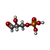


| #2: Chemical | ChemComp-G3H /  Mass: 170.058 Da / Num. of mol.: 8 / Source method: obtained synthetically / Formula: C3H7O6P / Feature type: SUBJECT OF INVESTIGATION Mass: 170.058 Da / Num. of mol.: 8 / Source method: obtained synthetically / Formula: C3H7O6P / Feature type: SUBJECT OF INVESTIGATION#3: Chemical | ChemComp-PEG /  Mass: 106.120 Da / Num. of mol.: 8 / Source method: obtained synthetically / Formula: C4H10O3 Mass: 106.120 Da / Num. of mol.: 8 / Source method: obtained synthetically / Formula: C4H10O3#4: Chemical | ChemComp-NAD /  Mass: 663.425 Da / Num. of mol.: 8 / Source method: obtained synthetically / Formula: C21H27N7O14P2 / Feature type: SUBJECT OF INVESTIGATION / Comment: NAD*YM Mass: 663.425 Da / Num. of mol.: 8 / Source method: obtained synthetically / Formula: C21H27N7O14P2 / Feature type: SUBJECT OF INVESTIGATION / Comment: NAD*YM#5: Chemical | ChemComp-NA /  Mass: 22.990 Da / Num. of mol.: 8 / Source method: obtained synthetically / Formula: Na Mass: 22.990 Da / Num. of mol.: 8 / Source method: obtained synthetically / Formula: Na#6: Chemical | ChemComp-PGE / |  Mass: 150.173 Da / Num. of mol.: 1 / Source method: obtained synthetically / Formula: C6H14O4 Mass: 150.173 Da / Num. of mol.: 1 / Source method: obtained synthetically / Formula: C6H14O4#7: Chemical | ChemComp-CL / |  Mass: 35.453 Da / Num. of mol.: 1 / Source method: obtained synthetically / Formula: Cl Mass: 35.453 Da / Num. of mol.: 1 / Source method: obtained synthetically / Formula: Cl#8: Chemical |  Mass: 92.094 Da / Num. of mol.: 2 / Source method: obtained synthetically / Formula: C3H8O3 Mass: 92.094 Da / Num. of mol.: 2 / Source method: obtained synthetically / Formula: C3H8O3#9: Chemical |  Mass: 238.305 Da / Num. of mol.: 2 / Source method: obtained synthetically / Formula: C8H18N2O4S / Comment: pH buffer*YM Mass: 238.305 Da / Num. of mol.: 2 / Source method: obtained synthetically / Formula: C8H18N2O4S / Comment: pH buffer*YM#10: Water | ChemComp-HOH / | Mass: 18.015 Da / Num. of mol.: 1298 / Source method: isolated from a natural source / Formula: H2O |
|---|
-Details
| Has ligand of interest | Y |
|---|---|
| Has protein modification | N |
-Experimental details
-Experiment
| Experiment | Method: X-RAY DIFFRACTION / Number of used crystals: 1 |
|---|
- Sample preparation
| Crystal | Density Matthews: 2.06 Å3/Da / Density % sol: 40.15 % |
|---|---|
| Crystal grow | Temperature: 291 K / Method: vapor diffusion, sitting drop / pH: 7.5 Details: Berkeley H9: 25% PEG 4000, 0.10M HEPES pH 7.5, 10% iso-Propanol. NegoA.00617.a.B1.PS38018 at 8 mg/mL. cocrystallization with NAD and G3H, plate 20061 H9 drop 1, Puck: PSL-2203, Cryo: 80% ...Details: Berkeley H9: 25% PEG 4000, 0.10M HEPES pH 7.5, 10% iso-Propanol. NegoA.00617.a.B1.PS38018 at 8 mg/mL. cocrystallization with NAD and G3H, plate 20061 H9 drop 1, Puck: PSL-2203, Cryo: 80% crystallant + 20% PEG 200 |
-Data collection
| Diffraction | Mean temperature: 100 K / Serial crystal experiment: N |
|---|---|
| Diffraction source | Source: SYNCHROTRON / Site: NSLS-II / Beamline: 19-ID / Wavelength: 0.9786 Å |
| Detector | Type: DECTRIS EIGER2 XE 9M / Detector: PIXEL / Date: Jun 28, 2025 |
| Radiation | Monochromator: Double Crystal Si 111 / Protocol: SINGLE WAVELENGTH / Monochromatic (M) / Laue (L): M / Scattering type: x-ray |
| Radiation wavelength | Wavelength: 0.9786 Å / Relative weight: 1 |
| Reflection | Resolution: 1.91→49.27 Å / Num. obs: 184320 / % possible obs: 99.9 % / Redundancy: 6.9 % / CC1/2: 0.996 / Rmerge(I) obs: 0.178 / Rpim(I) all: 0.073 / Rrim(I) all: 0.192 / Χ2: 1.04 / Net I/σ(I): 9.8 / Num. measured all: 1270381 |
| Reflection shell | Resolution: 1.91→1.96 Å / % possible obs: 100 % / Redundancy: 7.1 % / Rmerge(I) obs: 1.309 / Num. measured all: 95836 / Num. unique obs: 13577 / CC1/2: 0.581 / Rpim(I) all: 0.527 / Rrim(I) all: 1.413 / Χ2: 0.95 / Net I/σ(I) obs: 1.6 |
- Processing
| Software |
| |||||||||||||||||||||||||||||||||||||||||||||||||||||||||||||||||||||||||||||||||||||||||||||||||||||||||||||||||||||||||||||||||||||||||||||||||||||||||||||||||||||||||||||||||||||||||||||||||||||||||||||||||||||||||||||||||
|---|---|---|---|---|---|---|---|---|---|---|---|---|---|---|---|---|---|---|---|---|---|---|---|---|---|---|---|---|---|---|---|---|---|---|---|---|---|---|---|---|---|---|---|---|---|---|---|---|---|---|---|---|---|---|---|---|---|---|---|---|---|---|---|---|---|---|---|---|---|---|---|---|---|---|---|---|---|---|---|---|---|---|---|---|---|---|---|---|---|---|---|---|---|---|---|---|---|---|---|---|---|---|---|---|---|---|---|---|---|---|---|---|---|---|---|---|---|---|---|---|---|---|---|---|---|---|---|---|---|---|---|---|---|---|---|---|---|---|---|---|---|---|---|---|---|---|---|---|---|---|---|---|---|---|---|---|---|---|---|---|---|---|---|---|---|---|---|---|---|---|---|---|---|---|---|---|---|---|---|---|---|---|---|---|---|---|---|---|---|---|---|---|---|---|---|---|---|---|---|---|---|---|---|---|---|---|---|---|---|---|---|---|---|---|---|---|---|---|---|---|---|---|---|---|---|---|
| Refinement | Method to determine structure: MOLECULAR REPLACEMENT / Resolution: 1.91→48.89 Å / SU ML: 0.19 / Cross valid method: FREE R-VALUE / σ(F): 1.35 / Phase error: 18.72 / Stereochemistry target values: ML
| |||||||||||||||||||||||||||||||||||||||||||||||||||||||||||||||||||||||||||||||||||||||||||||||||||||||||||||||||||||||||||||||||||||||||||||||||||||||||||||||||||||||||||||||||||||||||||||||||||||||||||||||||||||||||||||||||
| Solvent computation | Shrinkage radii: 0.9 Å / VDW probe radii: 1.1 Å / Solvent model: FLAT BULK SOLVENT MODEL | |||||||||||||||||||||||||||||||||||||||||||||||||||||||||||||||||||||||||||||||||||||||||||||||||||||||||||||||||||||||||||||||||||||||||||||||||||||||||||||||||||||||||||||||||||||||||||||||||||||||||||||||||||||||||||||||||
| Refinement step | Cycle: LAST / Resolution: 1.91→48.89 Å
| |||||||||||||||||||||||||||||||||||||||||||||||||||||||||||||||||||||||||||||||||||||||||||||||||||||||||||||||||||||||||||||||||||||||||||||||||||||||||||||||||||||||||||||||||||||||||||||||||||||||||||||||||||||||||||||||||
| Refine LS restraints |
| |||||||||||||||||||||||||||||||||||||||||||||||||||||||||||||||||||||||||||||||||||||||||||||||||||||||||||||||||||||||||||||||||||||||||||||||||||||||||||||||||||||||||||||||||||||||||||||||||||||||||||||||||||||||||||||||||
| LS refinement shell |
| |||||||||||||||||||||||||||||||||||||||||||||||||||||||||||||||||||||||||||||||||||||||||||||||||||||||||||||||||||||||||||||||||||||||||||||||||||||||||||||||||||||||||||||||||||||||||||||||||||||||||||||||||||||||||||||||||
| Refinement TLS params. | Method: refined / Refine-ID: X-RAY DIFFRACTION
| |||||||||||||||||||||||||||||||||||||||||||||||||||||||||||||||||||||||||||||||||||||||||||||||||||||||||||||||||||||||||||||||||||||||||||||||||||||||||||||||||||||||||||||||||||||||||||||||||||||||||||||||||||||||||||||||||
| Refinement TLS group |
|
