 Movie
Movie Controller

[English] 日本語
 Yorodumi
- PDB-9z5u: Cryo-EM structure of a soluble HCV E1E2 antigen in complex with A... -
+ Open data

- Basic information
| Entry | Database: PDB / ID: 9z5u | |||||||||||||||||||||||||||
|---|---|---|---|---|---|---|---|---|---|---|---|---|---|---|---|---|---|---|---|---|---|---|---|---|---|---|---|---|
| Title | Cryo-EM structure of a soluble HCV E1E2 antigen in complex with AT1211 fab | |||||||||||||||||||||||||||
 Components Components |
| |||||||||||||||||||||||||||
 Keywords Keywords | VIRAL PROTEIN / Hepatitis C / broadly neutralizing antibody / soluble antigen | |||||||||||||||||||||||||||
| Biological species |  Hepacivirus hominis Hepacivirus hominis Homo sapiens (human) Homo sapiens (human) | |||||||||||||||||||||||||||
| Method | ELECTRON MICROSCOPY / single particle reconstruction / cryo EM / Resolution: 3.06 Å | |||||||||||||||||||||||||||
 Authors Authors | Cannac, F. / Ward, A.B. | |||||||||||||||||||||||||||
| Funding support | 1items
| |||||||||||||||||||||||||||
 Citation Citation | Journal: Nat Commun / Year: 2026 Title: Structure-based design of stable recombinant hepatitis C virus E1E2 heterodimers. Authors: Joan Capella-Pujol / Fabian Mulder / Fabien Cannac / Stan Peters / Maddy L Newby / Meliawati Poniman / Ian Zon / Wouter Olijhoek / Tim Beaumont / Max Crispin / Andrew B Ward / Janke Schinkel ...Authors: Joan Capella-Pujol / Fabian Mulder / Fabien Cannac / Stan Peters / Maddy L Newby / Meliawati Poniman / Ian Zon / Wouter Olijhoek / Tim Beaumont / Max Crispin / Andrew B Ward / Janke Schinkel / Rogier W Sanders / Kwinten Sliepen /    Abstract: Hepatitis C virus (HCV) affects 47 million people and causes 239,000 deaths annually, yet no vaccine is on the horizon. Generating a stable native-like mimic of the envelope complex E1E2, the only ...Hepatitis C virus (HCV) affects 47 million people and causes 239,000 deaths annually, yet no vaccine is on the horizon. Generating a stable native-like mimic of the envelope complex E1E2, the only target for known neutralizing antibodies, is an important aim for HCV vaccine development. Starting from a recombinant E1E2 design that utilizes a leucine zipper for proper folding, we used an iterative structure-based design approach to engineer antigens with at least 100-fold stronger binding to conformational antibodies and increased thermal stability. These new E1E2 designs facilitate production of native-like E1E2 antigens based on strains from different HCV genotypes and enable the generation of a recombinant E1E2 antigen design that lacks the immunogenic leucine zipper. A cryo-EM structure of one of the stabilized E1E2 antigens in complex with neutralizing antibody AT1211 provides atomic-level insights into an atypical epitope on the E2 subunit. Finally, immunogenicity studies in rabbits with adjuvanted E1E2 proteins show that immunogen stabilization alone does do not enhance serum neutralization breadth, but that removing the leucine zipper does increase homologous serum neutralization. | |||||||||||||||||||||||||||
| History |
|
- Structure visualization
| Structure viewer | Molecule:  MolmilJmol/JSmol MolmilJmol/JSmol |
|---|
- Downloads & links
-Download
| PDBx/mmCIF format | 9z5u.cif.gz | 112.8 KB | Display | PDBx/mmCIF format |
|---|---|---|---|---|
| PDB format | pdb9z5u.ent.gz | 75.5 KB | Display | PDB format |
| PDBx/mmJSON format | 9z5u.json.gz | Tree view | PDBx/mmJSON format | |
| Others |  Other downloads Other downloads |
-Validation report
| Arichive directory | https://data.pdbj.org/pub/pdb/validation_reports/z5/9z5uftp://data.pdbj.org/pub/pdb/validation_reports/z5/9z5u | HTTPS FTP |
|---|
-Related structure data

-Links
 PDBj
PDBj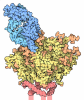


- Assembly
| Deposited unit | 
|
|---|---|
| 1 |
|
-Components
-Protein , 1 types, 1 molecules A
| #1: Protein | Mass: 64587.008 Da / Num. of mol.: 1 Source method: isolated from a genetically manipulated source Source: (gene. exp.) Hepacivirus hominis / Production host: Homo sapiens (human) |
|---|
-Antibody , 2 types, 2 molecules HL
| #2: Antibody | Mass: 24437.383 Da / Num. of mol.: 1 Source method: isolated from a genetically manipulated source Source: (gene. exp.) Homo sapiens (human) / Production host: Homo sapiens (human) |
|---|---|
| #3: Antibody | Mass: 23480.043 Da / Num. of mol.: 1 Source method: isolated from a genetically manipulated source Source: (gene. exp.) Homo sapiens (human) / Production host: Homo sapiens (human) |
-Sugars , 3 types, 4 molecules 
| #4: Polysaccharide | 2-acetamido-2-deoxy-beta-D-glucopyranose-(1-4)-2-acetamido-2-deoxy-beta-D-glucopyranose Source method: isolated from a genetically manipulated source |
|---|---|
| #5: Polysaccharide | beta-D-mannopyranose-(1-4)-2-acetamido-2-deoxy-beta-D-glucopyranose-(1-4)-2-acetamido-2-deoxy-beta- ...beta-D-mannopyranose-(1-4)-2-acetamido-2-deoxy-beta-D-glucopyranose-(1-4)-2-acetamido-2-deoxy-beta-D-glucopyranose Source method: isolated from a genetically manipulated source |
| #6: Sugar |  Type: D-saccharide, beta linking / Mass: 221.208 Da / Num. of mol.: 2 / Source method: obtained synthetically / Formula: C8H15NO6 Type: D-saccharide, beta linking / Mass: 221.208 Da / Num. of mol.: 2 / Source method: obtained synthetically / Formula: C8H15NO6 |
-Details
| Has ligand of interest | N |
|---|---|
| Has protein modification | Y |
-Experimental details
-Experiment
| Experiment | Method: ELECTRON MICROSCOPY |
|---|---|
| EM experiment | Aggregation state: PARTICLE / 3D reconstruction method: single particle reconstruction |
- Sample preparation
| Component | Name: Cryo-EM structure of a soluble HCV E1E2 antigen in complex with AT1211 fab Type: COMPLEX / Entity ID: #1-#3 / Source: MULTIPLE SOURCES |
|---|---|
| Molecular weight | Experimental value: NO |
| Source (natural) | Organism: Hepacivirus hominis |
| Source (recombinant) | Organism: Homo sapiens (human) |
| Buffer solution | pH: 7.4 |
| Specimen | Embedding applied: NO / Shadowing applied: NO / Staining applied: NO / Vitrification applied: YES |
| Specimen support | Grid material: COPPER / Grid mesh size: 300 divisions/in. / Grid type: Quantifoil R1.2/1.3 |
| Vitrification | Instrument: FEI VITROBOT MARK IV / Cryogen name: ETHANE / Humidity: 100 % / Chamber temperature: 277 K |
- Electron microscopy imaging
| Microscopy | Model: TFS GLACIOS |
|---|---|
| Electron gun | Electron source:  FIELD EMISSION GUN / Accelerating voltage: 200 kV / Illumination mode: FLOOD BEAM FIELD EMISSION GUN / Accelerating voltage: 200 kV / Illumination mode: FLOOD BEAM |
| Electron lens | Mode: BRIGHT FIELD / Nominal magnification: 190000 X / Nominal defocus max: 1600 nm / Nominal defocus min: 600 nm / Cs: 2.7 mm / C2 aperture diameter: 20 µm / Alignment procedure: COMA FREE |
| Specimen holder | Cryogen: NITROGEN / Specimen holder model: FEI TITAN KRIOS AUTOGRID HOLDER |
| Image recording | Average exposure time: 6.2 sec. / Electron dose: 60 e/Å2 / Film or detector model: TFS FALCON 4i (4k x 4k) / Num. of grids imaged: 1 / Num. of real images: 1827 |
- Processing
| EM software |
| ||||||||||||||||||||||||
|---|---|---|---|---|---|---|---|---|---|---|---|---|---|---|---|---|---|---|---|---|---|---|---|---|---|
| CTF correction | Type: PHASE FLIPPING AND AMPLITUDE CORRECTION | ||||||||||||||||||||||||
| Symmetry | Point symmetry: C1 (asymmetric) | ||||||||||||||||||||||||
| 3D reconstruction | Resolution: 3.06 Å / Resolution method: FSC 0.143 CUT-OFF / Num. of particles: 164770 / Symmetry type: POINT | ||||||||||||||||||||||||
| Refinement | Highest resolution: 3.06 Å Stereochemistry target values: REAL-SPACE (WEIGHTED MAP SUM AT ATOM CENTERS) | ||||||||||||||||||||||||
| Refine LS restraints |
|
