 Movie
Movie Controller

[English] 日本語
 Yorodumi
- PDB-9yup: Crystal structure of PprA S-F-S tetramer from Deinococcus radiodurans -
+ Open data

- Basic information
| Entry | Database: PDB / ID: 9yup | ||||||
|---|---|---|---|---|---|---|---|
| Title | Crystal structure of PprA S-F-S tetramer from Deinococcus radiodurans | ||||||
 Components Components | DNA repair protein PprA | ||||||
 Keywords Keywords | DNA BINDING PROTEIN / PprA / Deinococcus / Deinococcus radiodurans / D. radiodurans / DNA repair / Genome reassembly / self-assembly / protein filament | ||||||
| Function / homology | cellular response to desiccation / cellular response to gamma radiation / double-strand break repair via nonhomologous end joining / double-stranded DNA binding / damaged DNA binding / DNA repair / CITRATE ANION / DNA repair protein PprA Function and homology information Function and homology information | ||||||
| Biological species |  Deinococcus radiodurans (radioresistant) Deinococcus radiodurans (radioresistant) | ||||||
| Method |  X-RAY DIFFRACTION / SYNCHROTRON / SAD / Resolution: 2.07 Å X-RAY DIFFRACTION / SYNCHROTRON / SAD / Resolution: 2.07 Å | ||||||
 Authors Authors | Szabla, R. / Junop, M.S. | ||||||
| Funding support |  Canada, 1items Canada, 1items
| ||||||
 Citation Citation | Journal: To Be Published Title: Self-assembly of PprA from D.radiodurans Authors: Szabla, R. / Junop, M.S. | ||||||
| History |
|
- Structure visualization
| Structure viewer | Molecule: MolmilJmol/JSmol |
|---|
- Downloads & links
-Download
| PDBx/mmCIF format | 9yup.cif.gz | 396.9 KB | Display | PDBx/mmCIF format |
|---|---|---|---|---|
| PDB format | pdb9yup.ent.gz | 319.9 KB | Display | PDB format |
| PDBx/mmJSON format | 9yup.json.gz | Tree view | PDBx/mmJSON format | |
| Others |  Other downloads Other downloads |
-Validation report
| Summary document | 9yup_validation.pdf.gz | 493.4 KB | Display | wwPDB validaton report |
|---|---|---|---|---|
| Full document | 9yup_full_validation.pdf.gz | 515.1 KB | Display | |
| Data in XML | 9yup_validation.xml.gz | 47.6 KB | Display | |
| Data in CIF | 9yup_validation.cif.gz | 61.5 KB | Display | |
| Arichive directory | https://data.pdbj.org/pub/pdb/validation_reports/yu/9yupftp://data.pdbj.org/pub/pdb/validation_reports/yu/9yup | HTTPS FTP |
-Related structure data
| Related structure data | 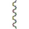 9om8C  9or6C  9yi3C  9yl4C C: citing same article ( |
|---|---|
| Similar structure data | |
| Experimental dataset #1 | Data reference: 10.5281/zenodo.17420657 / Data set type: diffraction image data / Metadata reference: 10.5281/zenodo.17420657 |
-Links
 PDBj
PDBj


- Assembly
| Deposited unit | 
| ||||||||||||
|---|---|---|---|---|---|---|---|---|---|---|---|---|---|
| 1 |
| ||||||||||||
| Unit cell |
|
-Components
| #1: Protein | Mass: 33602.750 Da / Num. of mol.: 4 / Mutation: D180K, D184K Source method: isolated from a genetically manipulated source Details: 1-8 deletion of PprA from D.radiodurans with D180K/D184K mutation and N-terminal poly-His fusion tag separated by a TEV protease site Source: (gene. exp.) Deinococcus radiodurans (radioresistant)Strain: R1 / Gene: pprA, DR_A0346 / Plasmid: pMJ5671 Details (production host): pDEST-527-based expression plasmid Production host: #2: Chemical |   Mass: 189.100 Da / Num. of mol.: 3 / Source method: obtained synthetically / Formula: C6H5O7 Mass: 189.100 Da / Num. of mol.: 3 / Source method: obtained synthetically / Formula: C6H5O7#3: Water | ChemComp-HOH / |  Mass: 18.015 Da / Num. of mol.: 171 / Source method: isolated from a natural source / Formula: H2O Mass: 18.015 Da / Num. of mol.: 171 / Source method: isolated from a natural source / Formula: H2OHas ligand of interest | N | Has protein modification | Y | |
|---|
-Experimental details
-Experiment
| Experiment | Method: X-RAY DIFFRACTION / Number of used crystals: 1 |
|---|
- Sample preparation
| Crystal | Density Matthews: 3.08 Å3/Da / Density % sol: 60.1 % |
|---|---|
| Crystal grow | Temperature: 293.15 K / Method: vapor diffusion, hanging drop / pH: 7.5 Details: 1.0 ul of protein solution was mixed with 1.0 uL of crystallization solution and hung upside-down in a sealed chamber containing 1mL of well solution. | Protein solution: 2.4 mg/mL PprA (73 ...Details: 1.0 ul of protein solution was mixed with 1.0 uL of crystallization solution and hung upside-down in a sealed chamber containing 1mL of well solution. | Protein solution: 2.4 mg/mL PprA (73 uM), 150mM KCl, 20mM Tris, pH 7.5 | Crystallization solution: 400 mM Lithium citrate, 20% (w/v) PEG 3350 | Well solution: 1.5 M Ammonium sulfate Temp details: Temperature-controlled incubator |
-Data collection
| Diffraction | Mean temperature: 100 K / Ambient temp details: Nitrogen cryo-stream / Serial crystal experiment: N |
|---|---|
| Diffraction source | Source: SYNCHROTRON / Site: APS  / Beamline: 17-ID / Wavelength: 0.97625 Å / Beamline: 17-ID / Wavelength: 0.97625 Å |
| Detector | Type: DECTRIS PILATUS 6M / Detector: PIXEL / Date: Apr 24, 2017 / Details: IMCA-CAT default optics (April 2017) |
| Radiation | Monochromator: IMCA-CAT default optics (April 2017) / Protocol: SINGLE WAVELENGTH / Monochromatic (M) / Laue (L): M / Scattering type: x-ray |
| Radiation wavelength | Wavelength: 0.97625 Å / Relative weight: 1 |
| Reflection | Resolution: 2.068→92.207 Å / Num. obs: 105944 / % possible obs: 91.9 % / Redundancy: 10.7 % / Biso Wilson estimate: 36.58 Å2 / CC1/2: 0.996 / Rmerge(I) obs: 0.204 / Rpim(I) all: 0.067 / Rrim(I) all: 0.223 / Net I/σ(I): 9.7 |
| Reflection shell | Resolution: 2.068→2.409 Å / Redundancy: 9.8 % / Rmerge(I) obs: 1.775 / Mean I/σ(I) obs: 1.9 / Num. unique obs: 5298 / CC1/2: 0.588 / Rpim(I) all: 0.605 / Rrim(I) all: 1.936 / % possible all: 63.2 |
- Processing
| Software |
| |||||||||||||||||||||||||||||||||||||||||||||||||||||||||||||||||||||||||||||||||||||||||||||||||||||||||||||||||||||||||||||||||||||||||||||||||||||||||||||||||||||||||||||||||||||||||||||||||||||||||||||||||||||||||
|---|---|---|---|---|---|---|---|---|---|---|---|---|---|---|---|---|---|---|---|---|---|---|---|---|---|---|---|---|---|---|---|---|---|---|---|---|---|---|---|---|---|---|---|---|---|---|---|---|---|---|---|---|---|---|---|---|---|---|---|---|---|---|---|---|---|---|---|---|---|---|---|---|---|---|---|---|---|---|---|---|---|---|---|---|---|---|---|---|---|---|---|---|---|---|---|---|---|---|---|---|---|---|---|---|---|---|---|---|---|---|---|---|---|---|---|---|---|---|---|---|---|---|---|---|---|---|---|---|---|---|---|---|---|---|---|---|---|---|---|---|---|---|---|---|---|---|---|---|---|---|---|---|---|---|---|---|---|---|---|---|---|---|---|---|---|---|---|---|---|---|---|---|---|---|---|---|---|---|---|---|---|---|---|---|---|---|---|---|---|---|---|---|---|---|---|---|---|---|---|---|---|---|---|---|---|---|---|---|---|---|---|---|---|---|---|---|---|---|
| Refinement | Method to determine structure: SAD / Resolution: 2.07→41.65 Å / SU ML: 0.1821 / Cross valid method: FREE R-VALUE / σ(F): 1.33 / Phase error: 31.3904 Stereochemistry target values: GeoStd + Monomer Library + CDL v1.2
| |||||||||||||||||||||||||||||||||||||||||||||||||||||||||||||||||||||||||||||||||||||||||||||||||||||||||||||||||||||||||||||||||||||||||||||||||||||||||||||||||||||||||||||||||||||||||||||||||||||||||||||||||||||||||
| Solvent computation | Shrinkage radii: 0.9 Å / VDW probe radii: 1.1 Å / Solvent model: FLAT BULK SOLVENT MODEL | |||||||||||||||||||||||||||||||||||||||||||||||||||||||||||||||||||||||||||||||||||||||||||||||||||||||||||||||||||||||||||||||||||||||||||||||||||||||||||||||||||||||||||||||||||||||||||||||||||||||||||||||||||||||||
| Displacement parameters | Biso mean: 58.76 Å2 | |||||||||||||||||||||||||||||||||||||||||||||||||||||||||||||||||||||||||||||||||||||||||||||||||||||||||||||||||||||||||||||||||||||||||||||||||||||||||||||||||||||||||||||||||||||||||||||||||||||||||||||||||||||||||
| Refinement step | Cycle: LAST / Resolution: 2.07→41.65 Å
| |||||||||||||||||||||||||||||||||||||||||||||||||||||||||||||||||||||||||||||||||||||||||||||||||||||||||||||||||||||||||||||||||||||||||||||||||||||||||||||||||||||||||||||||||||||||||||||||||||||||||||||||||||||||||
| Refine LS restraints |
| |||||||||||||||||||||||||||||||||||||||||||||||||||||||||||||||||||||||||||||||||||||||||||||||||||||||||||||||||||||||||||||||||||||||||||||||||||||||||||||||||||||||||||||||||||||||||||||||||||||||||||||||||||||||||
| LS refinement shell |
|
