 Movie
Movie Controller

[English] 日本語
 Yorodumi
- PDB-9ylb: Cryo-EM structure of single-loaded human UBA6-UBE2Z/FAT10(a) aden... -
+ Open data

- Basic information
| Entry | Database: PDB / ID: 9ylb | |||||||||
|---|---|---|---|---|---|---|---|---|---|---|
| Title | Cryo-EM structure of single-loaded human UBA6-UBE2Z/FAT10(a) adenylate complex. | |||||||||
 Components Components |
| |||||||||
 Keywords Keywords | TRANSFERASE/Ligase / E1 E2 enzymes / TRANSFERASE / TRANSFERASE-Ligase complex | |||||||||
| Function / homology |  Function and homology information Function and homology informationFAT10 activating enzyme activity / E1 ubiquitin-activating enzyme / ubiquitin activating enzyme activity / E2 ubiquitin-conjugating enzyme / proteasome binding / response to stress / ubiquitin conjugating enzyme activity / response to tumor necrosis factor / Dengue Virus Attachment and Entry / Synthesis of active ubiquitin: roles of E1 and E2 enzymes ...FAT10 activating enzyme activity / E1 ubiquitin-activating enzyme / ubiquitin activating enzyme activity / E2 ubiquitin-conjugating enzyme / proteasome binding / response to stress / ubiquitin conjugating enzyme activity / response to tumor necrosis factor / Dengue Virus Attachment and Entry / Synthesis of active ubiquitin: roles of E1 and E2 enzymes / Antigen processing: Ubiquitination & Proteasome degradation / ubiquitin-dependent protein catabolic process / molecular adaptor activity / protein ubiquitination / positive regulation of apoptotic process / apoptotic process / DNA damage response / negative regulation of apoptotic process / metal ion binding / nucleoplasm / ATP binding / nucleus / cytosol / cytoplasm Similarity search - Function | |||||||||
| Biological species |  Homo sapiens (human) Homo sapiens (human) | |||||||||
| Method | ELECTRON MICROSCOPY / single particle reconstruction / cryo EM / Resolution: 3.24 Å | |||||||||
 Authors Authors | Jia, L. / Ruben, E.A. / Nayak, D. / Bury, P.S. / Nayak, A. / Wasmuth, E.V. / Olsen, S.K. | |||||||||
| Funding support |  United States, 1items United States, 1items
| |||||||||
 Citation Citation | Journal: Nat Commun / Year: 2026 Title: Cryo-EM structures of UBA6 reveal mechanisms of E1-E2 specificity and dual FAT10/ubiquitin thioester transfer. Authors: Digant Nayak / Lijia Jia / Priscila Dos Santos Bury / Eliza A Ruben / Ankita Shukla / Anindita Nayak / Caleb M Stratton / Pirouz Ebadi / Hee Cho / Anna A Tumanova / Joyce T Varughese / ...Authors: Digant Nayak / Lijia Jia / Priscila Dos Santos Bury / Eliza A Ruben / Ankita Shukla / Anindita Nayak / Caleb M Stratton / Pirouz Ebadi / Hee Cho / Anna A Tumanova / Joyce T Varughese / Lingmin Yuan / Fei Gao / Kristin E Cano / Christopher Davies / Patrick Sung / Michaela U Gack / Elizabeth V Wasmuth / Shaun K Olsen / Abstract: UBA1 and UBA6 define parallel ubiquitin (Ub) activation systems that perform non-overlapping roles in Ub and ubiquitin-like protein (Ubl) signaling. Whereas UBA1 supports the canonical Ub pathway, ...UBA1 and UBA6 define parallel ubiquitin (Ub) activation systems that perform non-overlapping roles in Ub and ubiquitin-like protein (Ubl) signaling. Whereas UBA1 supports the canonical Ub pathway, UBA6 also activates the Ubl FAT10, linking Ub signaling to immune-regulated proteostasis. In addition to selective Ub/Ubl activation, UBA1 and UBA6 engage distinct sets of E2s, yet how these enzymes achieve selective E2 engagement has remained unclear. Using chemical trapping and high-resolution cryo-EM, we determine four structures of UBA6-E2 complexes representing the thioester-transfer step with either FAT10 or Ub, revealing how this E1 distinguishes its cognate partners. UBA6 achieves E2 specificity through coordinated contributions of the UFD and SCCH domains, a dual-domain mechanism that contrasts with the UFD-dominated selectivity of UBA1. The structures further show that an existing inositol hexakisphosphate (InsP₆)-binding site, unique to UBA6, stabilizes an expanded SCCH cleft that pre-organizes the enzyme for selective engagement of UBA6-specific E2s. These findings define principles for E1-E2 recognition and identify InsP₆ as a cofactor shaping specificity within the Ub-like conjugation network. | |||||||||
| History |
|
- Structure visualization
| Structure viewer | Molecule: MolmilJmol/JSmol |
|---|
- Downloads & links
-Download
| PDBx/mmCIF format | 9ylb.cif.gz | 287.7 KB | Display | PDBx/mmCIF format |
|---|---|---|---|---|
| PDB format | pdb9ylb.ent.gz | 221.8 KB | Display | PDB format |
| PDBx/mmJSON format | 9ylb.json.gz | Tree view | PDBx/mmJSON format | |
| Others |  Other downloads Other downloads |
-Validation report
| Arichive directory | https://data.pdbj.org/pub/pdb/validation_reports/yl/9ylbftp://data.pdbj.org/pub/pdb/validation_reports/yl/9ylb | HTTPS FTP |
|---|
-Related structure data
| Related structure data |  73079MC  9ykvC  9ykwC  9ylfC M: map data used to model this data C: citing same article ( |
|---|---|
| Similar structure data |


-Links
 PDBj
PDBj



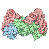
- Assembly
| Deposited unit | 
|
|---|---|
| 1 |
|
-Components
| #1: Protein | Mass: 114475.867 Da / Num. of mol.: 1 Source method: isolated from a genetically manipulated source Source: (gene. exp.) Homo sapiens (human) / Gene: UBA6, MOP4, UBE1L2 / Production host:  Trichoplusia ni (cabbage looper) / References: UniProt: A0AVT1, E1 ubiquitin-activating enzyme Trichoplusia ni (cabbage looper) / References: UniProt: A0AVT1, E1 ubiquitin-activating enzyme |
|---|---|
| #2: Protein | Mass: 29822.865 Da / Num. of mol.: 1 / Mutation: C100S,C261S,C263S, Y182A Source method: isolated from a genetically manipulated source Source: (gene. exp.) Homo sapiens (human) / Gene: UBE2Z, HOYS7 / Production host:   Escherichia coli (E. coli) Escherichia coli (E. coli)References: UniProt: Q9H832, E2 ubiquitin-conjugating enzyme |
| #3: Protein | Mass: 18476.471 Da / Num. of mol.: 1 / Mutation: C7T, C9T,C134L,C160S,C162S Source method: isolated from a genetically manipulated source Source: (gene. exp.) Homo sapiens (human) / Production host: Escherichia coli (E. coli) / References: UniProt: A0A1U9X8S9 |
| #4: Chemical | ChemComp-IHP /  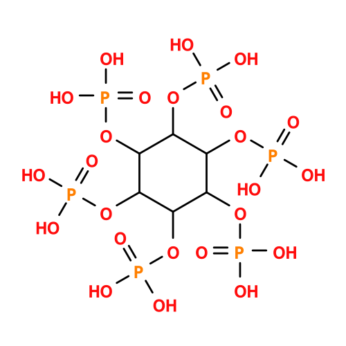 Mass: 660.035 Da / Num. of mol.: 1 / Source method: obtained synthetically / Formula: C6H18O24P6 Mass: 660.035 Da / Num. of mol.: 1 / Source method: obtained synthetically / Formula: C6H18O24P6 |
| #5: Chemical | ChemComp-AMP /  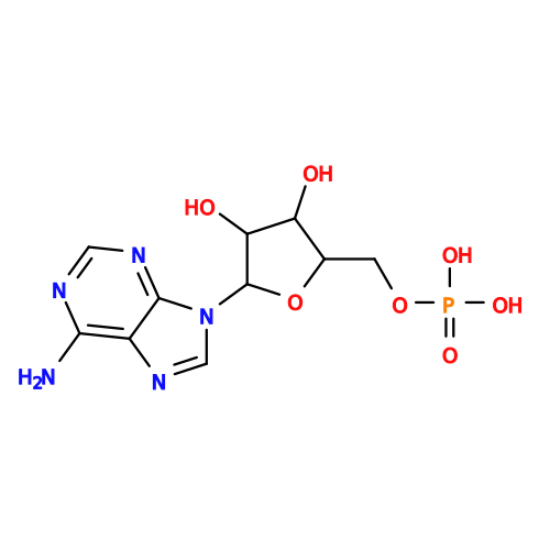 Mass: 347.221 Da / Num. of mol.: 1 / Source method: obtained synthetically / Formula: C10H14N5O7P / Feature type: SUBJECT OF INVESTIGATION / Comment: AMP*YM Mass: 347.221 Da / Num. of mol.: 1 / Source method: obtained synthetically / Formula: C10H14N5O7P / Feature type: SUBJECT OF INVESTIGATION / Comment: AMP*YM |
| Has ligand of interest | Y |
| Has protein modification | Y |
-Experimental details
-Experiment
| Experiment | Method: ELECTRON MICROSCOPY |
|---|---|
| EM experiment | Aggregation state: CELL / 3D reconstruction method: single particle reconstruction |
- Sample preparation
| Component | Name: Complex / Type: COMPLEX / Entity ID: #1-#3 / Source: RECOMBINANT |
|---|---|
| Source (natural) | Organism: Homo sapiens (human) |
| Source (recombinant) | Organism: Escherichia coli (E. coli) |
| Buffer solution | pH: 7.5 |
| Specimen | Embedding applied: NO / Shadowing applied: NO / Staining applied: NO / Vitrification applied: YES |
| Vitrification | Cryogen name: ETHANE |
- Electron microscopy imaging
| Experimental equipment |  Model: Titan Krios / Image courtesy: FEI Company |
|---|---|
| Microscopy | Model: TFS KRIOS |
| Electron gun | Electron source:  FIELD EMISSION GUN / Accelerating voltage: 300 kV / Illumination mode: FLOOD BEAM FIELD EMISSION GUN / Accelerating voltage: 300 kV / Illumination mode: FLOOD BEAM |
| Electron lens | Mode: BRIGHT FIELD / Nominal defocus max: 2500 nm / Nominal defocus min: 800 nm |
| Image recording | Electron dose: 53 e/Å2 / Film or detector model: GATAN K3 BIOCONTINUUM (6k x 4k) |
- Processing
| EM software |
| ||||||||||||||||||||||||
|---|---|---|---|---|---|---|---|---|---|---|---|---|---|---|---|---|---|---|---|---|---|---|---|---|---|
| CTF correction | Type: PHASE FLIPPING AND AMPLITUDE CORRECTION | ||||||||||||||||||||||||
| 3D reconstruction | Resolution: 3.24 Å / Resolution method: FSC 0.143 CUT-OFF / Num. of particles: 265430 / Symmetry type: POINT | ||||||||||||||||||||||||
| Refinement | Highest resolution: 3.24 Å Stereochemistry target values: REAL-SPACE (WEIGHTED MAP SUM AT ATOM CENTERS) | ||||||||||||||||||||||||
| Refine LS restraints |
|
