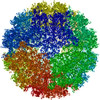
Movie
Movie Controller

[English] 日本語
 Yorodumi
- PDB-9yb1: Localized reconstruction of the asymmetric unit of SINV/EEEV at p... -
+ Open data

- Basic information
| Entry | Database: PDB / ID: 9yb1 | |||||||||||||||||||||||||||||||||
|---|---|---|---|---|---|---|---|---|---|---|---|---|---|---|---|---|---|---|---|---|---|---|---|---|---|---|---|---|---|---|---|---|---|---|
| Title | Localized reconstruction of the asymmetric unit of SINV/EEEV at pH 5.6. | |||||||||||||||||||||||||||||||||
 Components Components |
| |||||||||||||||||||||||||||||||||
 Keywords Keywords | VIRUS / Eastern Equine Encephalitis Virus / Cryo-EM / Single Particle Averaging / localized reconstruction / asymmetric unit / low pH back neutralization. | |||||||||||||||||||||||||||||||||
| Function / homology |  Function and homology information Function and homology informationT=4 icosahedral viral capsid / host cell cytoplasm / serine-type endopeptidase activity / fusion of virus membrane with host endosome membrane / symbiont entry into host cell / virion attachment to host cell / host cell nucleus / host cell plasma membrane / virion membrane / structural molecule activity ...T=4 icosahedral viral capsid / host cell cytoplasm / serine-type endopeptidase activity / fusion of virus membrane with host endosome membrane / symbiont entry into host cell / virion attachment to host cell / host cell nucleus / host cell plasma membrane / virion membrane / structural molecule activity / proteolysis / RNA binding Similarity search - Function | |||||||||||||||||||||||||||||||||
| Biological species |  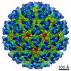 Eastern equine encephalitis virus Eastern equine encephalitis virus | |||||||||||||||||||||||||||||||||
| Method | ELECTRON MICROSCOPY / single particle reconstruction / cryo EM / Resolution: 4.7 Å | |||||||||||||||||||||||||||||||||
 Authors Authors | Bandyopadhyay, A. / Klose, T. / Kuhn, R.J. | |||||||||||||||||||||||||||||||||
| Funding support |  United States, 1items United States, 1items
| |||||||||||||||||||||||||||||||||
 Citation Citation | Journal: Nat Commun / Year: 2026 Title: Asymmetric structural transitions in the icosahedral organization of Eastern equine encephalitis virus. Authors: Abhishek Bandyopadhyay / Lauren E Williamson / Cameron D Buchman / Thomas Klose / James E Crowe / Richard J Kuhn / Abstract: Delivery of the viral genome into host cells is a critical step in successful viral infection. Alphaviruses achieve this step by fusing the viral and endosomal membranes under acidic conditions. This ...Delivery of the viral genome into host cells is a critical step in successful viral infection. Alphaviruses achieve this step by fusing the viral and endosomal membranes under acidic conditions. This process requires significant structural changes in the alphavirus glycoprotein organization. Structural characterization of acidic pH-induced conformational changes in alphavirus virions has remained elusive due to the rapid, transient nature of these states, conformational heterogeneity, and particle aggregation. Antibody binding studies conducted at elevated temperatures or under acidic pH conditions have further revealed the presence of transitional epitopes that are inaccessible on alphaviruses at room temperature or neutral pH. In this report, we present structural snapshots of the conformational changes in the glycoproteins and nucleocapsid core of a prototypical alphavirus, Eastern equine encephalitis virus, caused by exposure to 40 °C or pH 5.6. These findings provide insights into the structural transitions that occur prior to viral fusion with the endosomal membrane. This approach has also allowed us to define the molecular basis for recognition of a pan-alphavirus epitope by a patient-derived human antibody. | |||||||||||||||||||||||||||||||||
| History |
|
- Structure visualization
| Structure viewer | Molecule: MolmilJmol/JSmol |
|---|
- Downloads & links
-Download
| PDBx/mmCIF format | 9yb1.cif.gz | 687.3 KB | Display | PDBx/mmCIF format |
|---|---|---|---|---|
| PDB format | pdb9yb1.ent.gz | 575.5 KB | Display | PDB format |
| PDBx/mmJSON format | 9yb1.json.gz | Tree view | PDBx/mmJSON format | |
| Others |  Other downloads Other downloads |
-Validation report
| Arichive directory | https://data.pdbj.org/pub/pdb/validation_reports/yb/9yb1ftp://data.pdbj.org/pub/pdb/validation_reports/yb/9yb1 | HTTPS FTP |
|---|
-Related structure data
| Related structure data |  72745MC  9pu2C  9yawC  9yaxC  9yazC  9yb2C  9yb3C  9yb4C M: map data used to model this data C: citing same article ( |
|---|---|
| Similar structure data |


-Links
 PDBj
PDBj
- Assembly
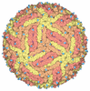
| Deposited unit | 
|
|---|---|
| 1 |
|
-Components
| #1: Protein | Mass: 47357.480 Da / Num. of mol.: 4 Source method: isolated from a genetically manipulated source Source: (gene. exp.) Eastern equine encephalitis virus / Strain: FL 93-939 / Gene: SP, F782_44616gpSP, F782_44619gpSP / Production host:  Mesocricetus auratus (golden hamster) / References: UniProt: W8RHT7 Mesocricetus auratus (golden hamster) / References: UniProt: W8RHT7#2: Protein | Mass: 17650.041 Da / Num. of mol.: 4 / Fragment: Capsid c-terminal domain Source method: isolated from a genetically manipulated source Source: (gene. exp.) Eastern equine encephalitis virus / Strain: FL 93-939 / Production host: Mesocricetus auratus (golden hamster) / References: UniProt: Q88793#3: Protein | Mass: 47046.953 Da / Num. of mol.: 4 / Fragment: UNP residues 325-744 Source method: isolated from a genetically manipulated source Source: (gene. exp.) Eastern equine encephalitis virus / Strain: FL 93-939 / Production host: Mesocricetus auratus (golden hamster) / References: UniProt: E9KXL2, togavirin#4: Polysaccharide | beta-D-mannopyranose-(1-4)-2-acetamido-2-deoxy-beta-D-glucopyranose-(1-4)-2-acetamido-2-deoxy-beta- ...beta-D-mannopyranose-(1-4)-2-acetamido-2-deoxy-beta-D-glucopyranose-(1-4)-2-acetamido-2-deoxy-beta-D-glucopyranose Source method: isolated from a genetically manipulated source #5: Sugar | ChemComp-NAG /   Type: D-saccharide, beta linking / Mass: 221.208 Da / Num. of mol.: 4 / Source method: obtained synthetically / Formula: C8H15NO6 Type: D-saccharide, beta linking / Mass: 221.208 Da / Num. of mol.: 4 / Source method: obtained synthetically / Formula: C8H15NO6Has ligand of interest | N | Has protein modification | Y | |
|---|
-Experimental details
-Experiment
| Experiment | Method: ELECTRON MICROSCOPY |
|---|---|
| EM experiment | Aggregation state: PARTICLE / 3D reconstruction method: single particle reconstruction |
- Sample preparation
| Component | Name: Eastern equine encephalitis virus / Type: VIRUS / Entity ID: #1-#3 / Source: RECOMBINANT |
|---|---|
| Molecular weight | Experimental value: NO |
| Source (natural) | Organism: Eastern equine encephalitis virus |
| Source (recombinant) | Organism:  Culiseta melanura (mosquito) Culiseta melanura (mosquito) |
| Details of virus | Empty: NO / Enveloped: YES / Isolate: STRAIN / Type: VIRION |
| Natural host | Organism: Culiseta melanura |
| Buffer solution | pH: 5.6 |
| Specimen | Conc.: 1 mg/ml / Embedding applied: NO / Shadowing applied: NO / Staining applied: NO / Vitrification applied: YES |
| Vitrification | Cryogen name: ETHANE / Humidity: 90 % / Chamber temperature: 298 K |
- Electron microscopy imaging
| Experimental equipment |  Model: Titan Krios / Image courtesy: FEI Company |
|---|---|
| Microscopy | Model: TFS KRIOS |
| Electron gun | Electron source:  FIELD EMISSION GUN / Accelerating voltage: 300 kV / Illumination mode: FLOOD BEAM FIELD EMISSION GUN / Accelerating voltage: 300 kV / Illumination mode: FLOOD BEAM |
| Electron lens | Mode: BRIGHT FIELD / Nominal defocus max: 2000 nm / Nominal defocus min: 600 nm / Alignment procedure: COMA FREE |
| Specimen holder | Cryogen: NITROGEN / Specimen holder model: FEI TITAN KRIOS AUTOGRID HOLDER |
| Image recording | Electron dose: 36.2 e/Å2 / Film or detector model: GATAN K3 BIOQUANTUM (6k x 4k) |
| EM imaging optics | Energyfilter name: GIF Bioquantum / Energyfilter slit width: 20 eV |
- Processing
| EM software |
| ||||||||||||||||||||||||||||||||||||||||
|---|---|---|---|---|---|---|---|---|---|---|---|---|---|---|---|---|---|---|---|---|---|---|---|---|---|---|---|---|---|---|---|---|---|---|---|---|---|---|---|---|---|
| CTF correction | Type: PHASE FLIPPING AND AMPLITUDE CORRECTION | ||||||||||||||||||||||||||||||||||||||||
| Symmetry | Point symmetry: C1 (asymmetric) | ||||||||||||||||||||||||||||||||||||||||
| 3D reconstruction | Resolution: 4.7 Å / Resolution method: FSC 0.143 CUT-OFF / Num. of particles: 265738 / Symmetry type: POINT | ||||||||||||||||||||||||||||||||||||||||
| Atomic model building | Protocol: OTHER | ||||||||||||||||||||||||||||||||||||||||
| Atomic model building | PDB-ID: 6MX4 Accession code: 6MX4 / Source name: PDB / Type: experimental model | ||||||||||||||||||||||||||||||||||||||||
| Refinement | Highest resolution: 4.7 Å Stereochemistry target values: REAL-SPACE (WEIGHTED MAP SUM AT ATOM CENTERS) |