 Movie
Movie Controller

[English] 日本語
 Yorodumi
- PDB-9y2f: Metabotropic Glutamate Receptor 7 in complex with ecto-domain of ... -
+ Open data

- Basic information
| Entry | Database: PDB / ID: 9y2f | ||||||||||||||||||||||||
|---|---|---|---|---|---|---|---|---|---|---|---|---|---|---|---|---|---|---|---|---|---|---|---|---|---|
| Title | Metabotropic Glutamate Receptor 7 in complex with ecto-domain of Extracellular Leucine Rich Repeat and Fibronectin Type III Domain Containing 2 | ||||||||||||||||||||||||
 Components Components |
| ||||||||||||||||||||||||
 Keywords Keywords | MEMBRANE PROTEIN / mGluR7 / ELFN2 / GPCR / glutamate / trans-synaptic | ||||||||||||||||||||||||
| Function / homology |  Function and homology information Function and homology informationnegative regulation of glutamate secretion / group III metabotropic glutamate receptor activity / adenylate cyclase-inhibiting G protein-coupled glutamate receptor signaling pathway / serine binding / synaptic membrane adhesion / G protein-coupled glutamate receptor signaling pathway / Class C/3 (Metabotropic glutamate/pheromone receptors) / glutamate receptor activity / presynaptic active zone / glutamate binding ...negative regulation of glutamate secretion / group III metabotropic glutamate receptor activity / adenylate cyclase-inhibiting G protein-coupled glutamate receptor signaling pathway / serine binding / synaptic membrane adhesion / G protein-coupled glutamate receptor signaling pathway / Class C/3 (Metabotropic glutamate/pheromone receptors) / glutamate receptor activity / presynaptic active zone / glutamate binding / protein phosphatase inhibitor activity / axon development / asymmetric synapse / regulation of synaptic transmission, glutamatergic / sensory perception of sound / adenylate cyclase inhibitor activity / dendritic shaft / establishment of protein localization / PDZ domain binding / postsynaptic density membrane / adenylate cyclase-inhibiting G protein-coupled receptor signaling pathway / signaling receptor activity / chemical synaptic transmission / cell cortex / G alpha (i) signalling events / signaling receptor complex / postsynaptic membrane / protein dimerization activity / axon / calcium ion binding / dendrite / : / membrane / plasma membrane Similarity search - Function | ||||||||||||||||||||||||
| Biological species |  Homo sapiens (human) Homo sapiens (human) | ||||||||||||||||||||||||
| Method | ELECTRON MICROSCOPY / single particle reconstruction / cryo EM / Resolution: 4.6 Å | ||||||||||||||||||||||||
 Authors Authors | Ludlam, W.G. / Chang, C.T. / Liauw, B.W. / Cho, H.J. / Sawh-Gopal, A. / Izard, T. / Bao, H. / Dunn, H.A. / Vafabakhsh, R. / Martemyanov, K.A. | ||||||||||||||||||||||||
| Funding support |  United States, 2items United States, 2items
| ||||||||||||||||||||||||
 Citation Citation | Journal: Sci Adv / Year: 2026 Title: Structural basis for modulation of group III mGlu receptors by transsynaptic interactions. Authors: William G Ludlam / Chu-Ting Chang / Kristina Cechova / Brandon W Liauw / Hwa-Jin Cho / Safoura Salar / Anjelique Sawh-Gopal / Afroza Parvin / Simrat K Dhaliwal / Simran K Dhaliwal / Tina ...Authors: William G Ludlam / Chu-Ting Chang / Kristina Cechova / Brandon W Liauw / Hwa-Jin Cho / Safoura Salar / Anjelique Sawh-Gopal / Afroza Parvin / Simrat K Dhaliwal / Simran K Dhaliwal / Tina Izard / Huan Bao / Anne M Brown / Henry A Dunn / Reza Vafabakhsh / Kirill A Martemyanov /  Abstract: Group III metabotropic glutamate receptors (mGluRs) are critical signaling molecules that regulate strength, homeostasis, and plasticity of glutamatergic synaptic signaling. These receptors are ...Group III metabotropic glutamate receptors (mGluRs) are critical signaling molecules that regulate strength, homeostasis, and plasticity of glutamatergic synaptic signaling. These receptors are engaged in transsynaptic interactions with extracellular leucine-rich repeat and fibronectin type III domain-containing (ELFN) cell adhesion proteins. ELFN proteins have been shown to play a critical role in regulation of activity and localization of mGluRs activity in vivo, yet the exact nature of their regulatory interaction has remained unknown. Here, we present a cryo-electron microscopy structure of the ELFN-mGluR complex. We identify a specific ELFN-binding pocket on mGluRs involved in its allosteric regulation through the network of residues affecting the orthosteric ligand binding site. We further uncover cooperativity whereby mGluR activation increases their association with ELFN proteins as a potential feedback mechanism to regulate synaptic strength. Last, we determine that disruption in mGluR-ELFN interaction is a recurring mechanism underlying several neurological conditions as we delineate their structure-functional etiology. | ||||||||||||||||||||||||
| History |
|
- Structure visualization
| Structure viewer | Molecule: MolmilJmol/JSmol |
|---|
- Downloads & links
-Download
| PDBx/mmCIF format | 9y2f.cif.gz | 273 KB | Display | PDBx/mmCIF format |
|---|---|---|---|---|
| PDB format | pdb9y2f.ent.gz | 206.5 KB | Display | PDB format |
| PDBx/mmJSON format | 9y2f.json.gz | Tree view | PDBx/mmJSON format | |
| Others |  Other downloads Other downloads |
-Validation report
| Arichive directory | https://data.pdbj.org/pub/pdb/validation_reports/y2/9y2fftp://data.pdbj.org/pub/pdb/validation_reports/y2/9y2f | HTTPS FTP |
|---|
-Related structure data
| Related structure data |  72409MC M: map data used to model this data C: citing same article ( |
|---|---|
| Similar structure data |
-Links
 PDBj
PDBj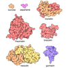
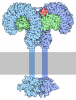
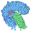
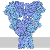
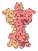

- Assembly
| Deposited unit | 
|
|---|---|
| 1 |
|
-Components
| #1: Protein | Mass: 43056.125 Da / Num. of mol.: 2 Source method: isolated from a genetically manipulated source Details: M1-A22 is cleaved off signal peptide. D23-G248 is leucine rich repeat domain. S249-T379 is not resolved in the structure. L380-Q385 is human rhinovirus C3 protease cut site sequence. Source: (gene. exp.) Homo sapiens (human) / Gene: ELFN2, KIAA1904, LRRC62, PPP1R29 / Production host: Homo sapiens (human) / References: UniProt: Q5R3F8#2: Protein | Mass: 97154.852 Da / Num. of mol.: 2 Source method: isolated from a genetically manipulated source Details: M1-G34 is cleaved signal peptide. Q35-M37 is not resolved in structure. Y38-S521 is Venus Flytrap domain. V522-R859 is not resolved in structure. L860-Q865 is human rhinovirus C3 protease cut site sequence. Source: (gene. exp.) Homo sapiens (human) / Gene: GRM7, GPRC1G, MGLUR7 / Production host: Homo sapiens (human) / References: UniProt: Q14831Has protein modification | Y | |
|---|
-Experimental details
-Experiment
| Experiment | Method: ELECTRON MICROSCOPY |
|---|---|
| EM experiment | Aggregation state: PARTICLE / 3D reconstruction method: single particle reconstruction |
- Sample preparation
| Component | Name: Complex of mGluR7 and ecto-domain of ELFN2 / Type: COMPLEX / Entity ID: all / Source: RECOMBINANT |
|---|---|
| Source (natural) | Organism: Homo sapiens (human) |
| Source (recombinant) | Organism: Homo sapiens (human) |
| Buffer solution | pH: 7.8 |
| Specimen | Embedding applied: NO / Shadowing applied: NO / Staining applied: NO / Vitrification applied: YES |
| Vitrification | Cryogen name: ETHANE |
- Electron microscopy imaging
| Microscopy | Model: JEOL CRYO ARM 300 |
|---|---|
| Electron gun | Electron source:  FIELD EMISSION GUN / Accelerating voltage: 300 kV / Illumination mode: FLOOD BEAM FIELD EMISSION GUN / Accelerating voltage: 300 kV / Illumination mode: FLOOD BEAM |
| Electron lens | Mode: BRIGHT FIELD / Nominal defocus max: 2000 nm / Nominal defocus min: 1000 nm / Cs: 2.7 mm |
| Image recording | Electron dose: 50 e/Å2 / Film or detector model: GATAN K3 (6k x 4k) |
- Processing
| EM software |
| ||||||||||||||||||||||||
|---|---|---|---|---|---|---|---|---|---|---|---|---|---|---|---|---|---|---|---|---|---|---|---|---|---|
| CTF correction | Type: PHASE FLIPPING AND AMPLITUDE CORRECTION | ||||||||||||||||||||||||
| 3D reconstruction | Resolution: 4.6 Å / Resolution method: FSC 0.143 CUT-OFF / Num. of particles: 28023 / Symmetry type: POINT | ||||||||||||||||||||||||
| Refinement | Highest resolution: 4.6 Å Stereochemistry target values: REAL-SPACE (WEIGHTED MAP SUM AT ATOM CENTERS) | ||||||||||||||||||||||||
| Refine LS restraints |
|
