 Movie
Movie Controller

[English] 日本語
 Yorodumi
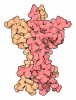
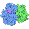
- PDB-9xw7: Cryo-EM structure of PI3Kalpha E545K mutation in complex with STX-478 -
+ Open data

- Basic information
| Entry | Database: PDB / ID: 9xw7 | ||||||||||||||||||
|---|---|---|---|---|---|---|---|---|---|---|---|---|---|---|---|---|---|---|---|
| Title | Cryo-EM structure of PI3Kalpha E545K mutation in complex with STX-478 | ||||||||||||||||||
 Components Components | Phosphatidylinositol 4,5-bisphosphate 3-kinase catalytic subunit alpha isoform | ||||||||||||||||||
 Keywords Keywords | STRUCTURAL PROTEIN / Phosphoinositide 3-kinase / drug target / ligand / binding pocket / hotspot mutation / allosteric inhbitor | ||||||||||||||||||
| Function / homology |  Function and homology information Function and homology informationresponse to muscle inactivity / regulation of actin filament organization / response to butyrate / negative regulation of actin filament depolymerization / IRS-mediated signalling / PI3K events in ERBB4 signaling / response to L-leucine / cellular response to hydrostatic pressure / Activated NTRK2 signals through PI3K / Activated NTRK3 signals through PI3K ...response to muscle inactivity / regulation of actin filament organization / response to butyrate / negative regulation of actin filament depolymerization / IRS-mediated signalling / PI3K events in ERBB4 signaling / response to L-leucine / cellular response to hydrostatic pressure / Activated NTRK2 signals through PI3K / Activated NTRK3 signals through PI3K / phosphatidylinositol 3-kinase complex, class IB / phosphatidylinositol 3-kinase complex / TORC2 signaling / Co-stimulation by ICOS / positive regulation of protein localization to membrane / Signaling by cytosolic FGFR1 fusion mutants / vasculature development / Nephrin family interactions / Signaling by LTK in cancer / 1-phosphatidylinositol-4-phosphate 3-kinase activity / Signaling by LTK / anoikis / phosphatidylinositol 3-kinase complex, class IA / MET activates PI3K/AKT signaling / PI3K/AKT activation / phosphatidylinositol-4,5-bisphosphate 3-kinase / 1-phosphatidylinositol-4,5-bisphosphate 3-kinase activity / relaxation of cardiac muscle / phosphatidylinositol 3-kinase / phosphatidylinositol-3-phosphate biosynthetic process / Signaling by ALK / cardiac muscle cell contraction / vascular endothelial growth factor signaling pathway / 1-phosphatidylinositol-3-kinase activity / Erythropoietin activates Phosphoinositide-3-kinase (PI3K) / PI-3K cascade:FGFR3 / negative regulation of macroautophagy / response to dexamethasone / PI-3K cascade:FGFR2 / PI-3K cascade:FGFR4 / PI-3K cascade:FGFR1 / phosphatidylinositol phosphate biosynthetic process / Synthesis of PIPs at the plasma membrane / positive regulation of protein kinase activity / response to muscle stretch / phosphatidylinositol-mediated signaling / RET signaling / negative regulation of anoikis / Interleukin-3, Interleukin-5 and GM-CSF signaling / PI3K events in ERBB2 signaling / insulin receptor substrate binding / PI3K Cascade / Role of LAT2/NTAL/LAB on calcium mobilization / RAC2 GTPase cycle / Interleukin receptor SHC signaling / intercalated disc / Role of phospholipids in phagocytosis / CD28 dependent PI3K/Akt signaling / positive regulation of TOR signaling / GAB1 signalosome / endothelial cell migration / protein kinase activator activity / positive regulation of lamellipodium assembly / cardiac muscle contraction / GPVI-mediated activation cascade / Signaling by PDGFRA transmembrane, juxtamembrane and kinase domain mutants / Signaling by PDGFRA extracellular domain mutants / insulin-like growth factor receptor signaling pathway / Signaling by FGFR4 in disease / Signaling by FLT3 ITD and TKD mutants / RAC1 GTPase cycle / Signaling by FGFR3 in disease / Tie2 Signaling / Signaling by FGFR2 in disease / Signaling by FLT3 fusion proteins / FLT3 Signaling / Signaling by FGFR1 in disease / positive regulation of smooth muscle cell proliferation / Downstream signal transduction / response to activity / Signaling by phosphorylated juxtamembrane, extracellular and kinase domain KIT mutants / phosphatidylinositol 3-kinase/protein kinase B signal transduction / Regulation of signaling by CBL / platelet activation / Constitutive Signaling by EGFRvIII / Signaling by SCF-KIT / Signaling by ERBB2 ECD mutants / Signaling by ERBB2 KD Mutants / VEGFA-VEGFR2 Pathway / insulin receptor signaling pathway / T cell receptor signaling pathway / epidermal growth factor receptor signaling pathway / Constitutive Signaling by Aberrant PI3K in Cancer / Signaling by CSF1 (M-CSF) in myeloid cells / actin cytoskeleton organization / cellular response to insulin stimulus / Signaling by ALK fusions and activated point mutants / DAP12 signaling / PIP3 activates AKT signaling / cell migration Similarity search - Function | ||||||||||||||||||
| Biological species |  Homo sapiens (human) Homo sapiens (human) | ||||||||||||||||||
| Method | ELECTRON MICROSCOPY / single particle reconstruction / cryo EM / Resolution: 3.01 Å | ||||||||||||||||||
 Authors Authors | Liu, X. / Chen, Y. / Li, G. / Chen, A. / Zhou, Q. / Wang, M.-W. | ||||||||||||||||||
| Funding support |  China, 5items China, 5items
| ||||||||||||||||||
 Citation Citation | Journal: Acta Pharm Sin B / Year: 2026 Title: Molecular architecture responsible for specific inhibition of oncogenic PI3K alpha mutants by RLY-2608 and STX-478 Authors: Liu, X. / Chen, Y. / Li, G. / Chen, A. / Zhou, Q. / Wang, M.W. | ||||||||||||||||||
| History |
|
- Structure visualization
| Structure viewer | Molecule: MolmilJmol/JSmol |
|---|
- Downloads & links
-Download
| PDBx/mmCIF format | 9xw7.cif.gz | 189.7 KB | Display | PDBx/mmCIF format |
|---|---|---|---|---|
| PDB format | pdb9xw7.ent.gz | 142 KB | Display | PDB format |
| PDBx/mmJSON format | 9xw7.json.gz | Tree view | PDBx/mmJSON format | |
| Others |  Other downloads Other downloads |
-Validation report
| Arichive directory | https://data.pdbj.org/pub/pdb/validation_reports/xw/9xw7ftp://data.pdbj.org/pub/pdb/validation_reports/xw/9xw7 | HTTPS FTP |
|---|
-Related structure data
| Related structure data |  67327MC  9xw5C  9xw8C M: map data used to model this data C: citing same article ( |
|---|---|
| Similar structure data |


-Links
 PDBj
PDBj











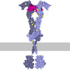
- Assembly
| Deposited unit | 
|
|---|---|
| 1 |
|
-Components
| #1: Protein | Mass: 127822.641 Da / Num. of mol.: 1 Source method: isolated from a genetically manipulated source Source: (gene. exp.) Homo sapiens (human) / Gene: PIK3CA / Production host:  Trichoplusia ni (cabbage looper) Trichoplusia ni (cabbage looper)References: UniProt: P42336, phosphatidylinositol 3-kinase, phosphatidylinositol-4,5-bisphosphate 3-kinase, non-specific serine/threonine protein kinase |
|---|---|
| #2: Chemical | ChemComp-ZWE /   Mass: 401.291 Da / Num. of mol.: 1 / Source method: obtained synthetically / Formula: C16H12F5N5O2 Mass: 401.291 Da / Num. of mol.: 1 / Source method: obtained synthetically / Formula: C16H12F5N5O2 |
| #3: Water | ChemComp-HOH /  Mass: 18.015 Da / Num. of mol.: 2 / Source method: isolated from a natural source / Formula: H2O Mass: 18.015 Da / Num. of mol.: 2 / Source method: isolated from a natural source / Formula: H2O |
| Has ligand of interest | N |
| Has protein modification | Y |
-Experimental details
-Experiment
| Experiment | Method: ELECTRON MICROSCOPY |
|---|---|
| EM experiment | Aggregation state: PARTICLE / 3D reconstruction method: single particle reconstruction |
- Sample preparation
| Component | Name: Human PI3Kalpha E542K in complex with STX-478 / Type: COMPLEX / Entity ID: #1 / Source: RECOMBINANT |
|---|---|
| Source (natural) | Organism: Homo sapiens (human) |
| Source (recombinant) | Organism: Trichoplusia ni (cabbage looper) |
| Buffer solution | pH: 7.6 |
| Specimen | Embedding applied: NO / Shadowing applied: NO / Staining applied: NO / Vitrification applied: YES |
| Vitrification | Cryogen name: ETHANE |
- Electron microscopy imaging
| Experimental equipment |  Model: Titan Krios / Image courtesy: FEI Company |
|---|---|
| Microscopy | Model: TFS KRIOS |
| Electron gun | Electron source: OTHER / Accelerating voltage: 300 kV / Illumination mode: OTHER |
| Electron lens | Mode: DIFFRACTION / Nominal defocus max: 2000 nm / Nominal defocus min: 1000 nm |
| Image recording | Electron dose: 50 e/Å2 / Film or detector model: GATAN K3 (6k x 4k) |
- Processing
| EM software |
| ||||||||||||||||||||||||
|---|---|---|---|---|---|---|---|---|---|---|---|---|---|---|---|---|---|---|---|---|---|---|---|---|---|
| CTF correction | Type: PHASE FLIPPING AND AMPLITUDE CORRECTION | ||||||||||||||||||||||||
| 3D reconstruction | Resolution: 3.01 Å / Resolution method: FSC 0.143 CUT-OFF / Num. of particles: 4873921 / Symmetry type: POINT | ||||||||||||||||||||||||
| Refinement | Highest resolution: 3.01 Å Stereochemistry target values: REAL-SPACE (WEIGHTED MAP SUM AT ATOM CENTERS) | ||||||||||||||||||||||||
| Refine LS restraints |
|
