 Movie
Movie Controller

[English] 日本語
 Yorodumi
- PDB-9xfy: Crystal structure of Class A beta-lactamase BlaA in complex with ... -
+ Open data

- Basic information
| Entry | Database: PDB / ID: 9xfy | |||||||||
|---|---|---|---|---|---|---|---|---|---|---|
| Title | Crystal structure of Class A beta-lactamase BlaA in complex with ertapenem (imine form) | |||||||||
 Components Components | Beta-lactamase | |||||||||
 Keywords Keywords | HYDROLASE / Imine tautomer / Carbapenemase / Acyl enzyme complex | |||||||||
| Function / homology |  Function and homology information Function and homology informationbeta-lactam antibiotic catabolic process / beta-lactamase activity / beta-lactamase / response to antibiotic Similarity search - Function | |||||||||
| Biological species |  Yersinia enterocolitica (bacteria) Yersinia enterocolitica (bacteria) | |||||||||
| Method |  X-RAY DIFFRACTION / MOLECULAR REPLACEMENT / Resolution: 1.7 Å X-RAY DIFFRACTION / MOLECULAR REPLACEMENT / Resolution: 1.7 Å | |||||||||
 Authors Authors | Bhattacharya, S. / Dhankhar, K. / Hazra, S. | |||||||||
| Funding support |  India, 2items India, 2items
| |||||||||
 Citation Citation | Journal: To Be Published Title: Crystal structure of Class A beta-lactamase BlaA in complex with ertapenem (imine form) Authors: Bhattacharya, S. / Dhankhar, K. / Hazra, S. | |||||||||
| History |
|
- Structure visualization
| Structure viewer | Molecule: MolmilJmol/JSmol |
|---|
- Downloads & links
-Download
| PDBx/mmCIF format | 9xfy.cif.gz | 74 KB | Display | PDBx/mmCIF format |
|---|---|---|---|---|
| PDB format | pdb9xfy.ent.gz | 51 KB | Display | PDB format |
| PDBx/mmJSON format | 9xfy.json.gz | Tree view | PDBx/mmJSON format | |
| Others |  Other downloads Other downloads |
-Validation report
| Arichive directory | https://data.pdbj.org/pub/pdb/validation_reports/xf/9xfyftp://data.pdbj.org/pub/pdb/validation_reports/xf/9xfy | HTTPS FTP |
|---|
-Related structure data
| Related structure data |  5e2eS S: Starting model for refinement |
|---|---|
| Similar structure data |
-Links
 PDBj
PDBj


- Assembly
| Deposited unit | 
| |||||||||
|---|---|---|---|---|---|---|---|---|---|---|
| 1 |
| |||||||||
| Unit cell |
| |||||||||
| Components on special symmetry positions |
|
-Components
| #1: Protein | Mass: 31966.578 Da / Num. of mol.: 1 Source method: isolated from a genetically manipulated source Source: (gene. exp.) Yersinia enterocolitica (bacteria) / Gene: blaA / Production host: | ||||||
|---|---|---|---|---|---|---|---|
| #2: Chemical | ChemComp-2RG / (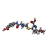  Mass: 477.531 Da / Num. of mol.: 1 / Source method: obtained synthetically / Formula: C22H27N3O7S / Feature type: SUBJECT OF INVESTIGATION / Comment: medication, antibiotic*YM Mass: 477.531 Da / Num. of mol.: 1 / Source method: obtained synthetically / Formula: C22H27N3O7S / Feature type: SUBJECT OF INVESTIGATION / Comment: medication, antibiotic*YM | ||||||
| #3: Chemical |   Mass: 94.971 Da / Num. of mol.: 3 / Source method: obtained synthetically / Formula: PO4 / Feature type: SUBJECT OF INVESTIGATION Mass: 94.971 Da / Num. of mol.: 3 / Source method: obtained synthetically / Formula: PO4 / Feature type: SUBJECT OF INVESTIGATION#4: Water | ChemComp-HOH / |  Mass: 18.015 Da / Num. of mol.: 212 / Source method: isolated from a natural source / Formula: H2O Mass: 18.015 Da / Num. of mol.: 212 / Source method: isolated from a natural source / Formula: H2OHas ligand of interest | Y | Has protein modification | Y | |
-Experimental details
-Experiment
| Experiment | Method: X-RAY DIFFRACTION / Number of used crystals: 1 |
|---|
- Sample preparation
| Crystal | Density Matthews: 2.29 Å3/Da / Density % sol: 45.85 % |
|---|---|
| Crystal grow | Temperature: 293 K / Method: vapor diffusion, sitting drop / pH: 8.5 Details: 0.1 M TRIS hydrochloride pH 8.5, 2.0 M Ammonium phosphate monobasic |
-Data collection
| Diffraction | Mean temperature: 100 K / Serial crystal experiment: N |
|---|---|
| Diffraction source | Source: ROTATING ANODE / Type: RIGAKU MICROMAX-007 HF / Wavelength: 1.54184 Å |
| Detector | Type: RIGAKU HyPix-6000HE / Detector: PIXEL / Date: Aug 17, 2022 |
| Radiation | Protocol: SINGLE WAVELENGTH / Monochromatic (M) / Laue (L): M / Scattering type: x-ray |
| Radiation wavelength | Wavelength: 1.54184 Å / Relative weight: 1 |
| Reflection | Resolution: 1.7→24.663 Å / Num. obs: 31443 / % possible obs: 99.8 % / Redundancy: 6.5 % / CC1/2: 0.989 / Rmerge(I) obs: 0.142 / Rpim(I) all: 0.078 / Rrim(I) all: 0.154 / Net I/σ(I): 7.4 |
| Reflection shell | Resolution: 1.7→1.73 Å / Rmerge(I) obs: 0.591 / Num. unique obs: 6673 / CC1/2: 0.809 / Rpim(I) all: 0.332 / Rrim(I) all: 0.682 |
- Processing
| Software |
| |||||||||||||||||||||||||||||||||||||||||||||||||||||||||||||||||||||||||||||||||||||||||||||||||||||||||||||||||||||||||||||||||||||||||||||||||||||||||||||||||||||||||||||||||||||||||||||||||||||||||||||||||||||||||||||||||||||||
|---|---|---|---|---|---|---|---|---|---|---|---|---|---|---|---|---|---|---|---|---|---|---|---|---|---|---|---|---|---|---|---|---|---|---|---|---|---|---|---|---|---|---|---|---|---|---|---|---|---|---|---|---|---|---|---|---|---|---|---|---|---|---|---|---|---|---|---|---|---|---|---|---|---|---|---|---|---|---|---|---|---|---|---|---|---|---|---|---|---|---|---|---|---|---|---|---|---|---|---|---|---|---|---|---|---|---|---|---|---|---|---|---|---|---|---|---|---|---|---|---|---|---|---|---|---|---|---|---|---|---|---|---|---|---|---|---|---|---|---|---|---|---|---|---|---|---|---|---|---|---|---|---|---|---|---|---|---|---|---|---|---|---|---|---|---|---|---|---|---|---|---|---|---|---|---|---|---|---|---|---|---|---|---|---|---|---|---|---|---|---|---|---|---|---|---|---|---|---|---|---|---|---|---|---|---|---|---|---|---|---|---|---|---|---|---|---|---|---|---|---|---|---|---|---|---|---|---|---|---|---|---|---|
| Refinement | Method to determine structure: MOLECULAR REPLACEMENT Starting model: 5E2E Resolution: 1.7→24.663 Å / Cor.coef. Fo:Fc: 0.946 / Cor.coef. Fo:Fc free: 0.922 / SU B: 2.508 / SU ML: 0.082 / Cross valid method: FREE R-VALUE / ESU R: 0.111 / ESU R Free: 0.11
| |||||||||||||||||||||||||||||||||||||||||||||||||||||||||||||||||||||||||||||||||||||||||||||||||||||||||||||||||||||||||||||||||||||||||||||||||||||||||||||||||||||||||||||||||||||||||||||||||||||||||||||||||||||||||||||||||||||||
| Solvent computation | Ion probe radii: 0.8 Å / Shrinkage radii: 0.8 Å / VDW probe radii: 1.2 Å / Solvent model: MASK BULK SOLVENT | |||||||||||||||||||||||||||||||||||||||||||||||||||||||||||||||||||||||||||||||||||||||||||||||||||||||||||||||||||||||||||||||||||||||||||||||||||||||||||||||||||||||||||||||||||||||||||||||||||||||||||||||||||||||||||||||||||||||
| Displacement parameters | Biso mean: 21.318 Å2
| |||||||||||||||||||||||||||||||||||||||||||||||||||||||||||||||||||||||||||||||||||||||||||||||||||||||||||||||||||||||||||||||||||||||||||||||||||||||||||||||||||||||||||||||||||||||||||||||||||||||||||||||||||||||||||||||||||||||
| Refinement step | Cycle: LAST / Resolution: 1.7→24.663 Å
| |||||||||||||||||||||||||||||||||||||||||||||||||||||||||||||||||||||||||||||||||||||||||||||||||||||||||||||||||||||||||||||||||||||||||||||||||||||||||||||||||||||||||||||||||||||||||||||||||||||||||||||||||||||||||||||||||||||||
| Refine LS restraints |
| |||||||||||||||||||||||||||||||||||||||||||||||||||||||||||||||||||||||||||||||||||||||||||||||||||||||||||||||||||||||||||||||||||||||||||||||||||||||||||||||||||||||||||||||||||||||||||||||||||||||||||||||||||||||||||||||||||||||
| LS refinement shell | Refine-ID: X-RAY DIFFRACTION / Total num. of bins used: 20
|
