 Movie
Movie Controller

[English] 日本語
 Yorodumi
- PDB-9x6m: Crystal structure of Klebsiella oxytoca ribitol dehydrogenase in ... -
+ Open data

- Basic information
| Entry | Database: PDB / ID: 9x6m | ||||||
|---|---|---|---|---|---|---|---|
| Title | Crystal structure of Klebsiella oxytoca ribitol dehydrogenase in complex with D-allose | ||||||
 Components Components | Ribitol 2-dehydrogenase | ||||||
 Keywords Keywords | OXIDOREDUCTASE / Rossmann fold / dehydrogenase | ||||||
| Function / homology | short chain dehydrogenase / Short-chain dehydrogenase/reductase, conserved site / Short-chain dehydrogenases/reductases family signature. / Short-chain dehydrogenase/reductase SDR / oxidoreductase activity / NAD(P)-binding domain superfamily / beta-D-allopyranose / Ribitol 2-dehydrogenase Function and homology information Function and homology information | ||||||
| Biological species |  Klebsiella oxytoca (bacteria) Klebsiella oxytoca (bacteria) | ||||||
| Method |  X-RAY DIFFRACTION / SYNCHROTRON / MOLECULAR REPLACEMENT / Resolution: 1.73 Å X-RAY DIFFRACTION / SYNCHROTRON / MOLECULAR REPLACEMENT / Resolution: 1.73 Å | ||||||
 Authors Authors | Yoshida, H. / Yoshihara, A. | ||||||
| Funding support |  Japan, 1items Japan, 1items
| ||||||
 Citation Citation | Journal: Febs Lett. / Year: 2026 Title: Crystal structures of Klebsiella oxytoca ribitol dehydrogenase in complex with NAD + , d-allose, or d-allulose reveal insight into substrate recognition. Authors: Yoshida, H. / Matsumoto, M. / Yamamoto, N. / Yoshihara, A. / Izumori, K. / Kamitori, S. | ||||||
| History |
|
- Structure visualization
| Structure viewer | Molecule: MolmilJmol/JSmol |
|---|
- Downloads & links
-Download
| PDBx/mmCIF format | 9x6m.cif.gz | 526.6 KB | Display | PDBx/mmCIF format |
|---|---|---|---|---|
| PDB format | pdb9x6m.ent.gz | 442.2 KB | Display | PDB format |
| PDBx/mmJSON format | 9x6m.json.gz | Tree view | PDBx/mmJSON format | |
| Others |  Other downloads Other downloads |
-Validation report
| Arichive directory | https://data.pdbj.org/pub/pdb/validation_reports/x6/9x6mftp://data.pdbj.org/pub/pdb/validation_reports/x6/9x6m | HTTPS FTP |
|---|
-Related structure data


-Links
 PDBj
PDBj


- Assembly
| Deposited unit | 
| ||||||||
|---|---|---|---|---|---|---|---|---|---|
| 1 |
| ||||||||
| Unit cell |
|
-Components
| #1: Protein | Mass: 28532.748 Da / Num. of mol.: 4 Source method: isolated from a genetically manipulated source Details: This is a His-tagged recombinant protein containing 21 residues (MGSSHHHHHHSSGLVPRGSHS ) at the N-terminus. A few residues are missing their side chains due to unclear electron density maps. ...Details: This is a His-tagged recombinant protein containing 21 residues (MGSSHHHHHHSSGLVPRGSHS ) at the N-terminus. A few residues are missing their side chains due to unclear electron density maps. They are refined as Ala but not mutated. Source: (gene. exp.) Klebsiella oxytoca (bacteria) / Gene: DET57_113163 / Plasmid: pET15b / Production host: #2: Sugar | 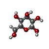  Type: D-saccharide, beta linking / Mass: 180.156 Da / Num. of mol.: 3 / Source method: obtained synthetically / Formula: C6H12O6 / Feature type: SUBJECT OF INVESTIGATION Type: D-saccharide, beta linking / Mass: 180.156 Da / Num. of mol.: 3 / Source method: obtained synthetically / Formula: C6H12O6 / Feature type: SUBJECT OF INVESTIGATION#3: Water | ChemComp-HOH / |  Mass: 18.015 Da / Num. of mol.: 669 / Source method: isolated from a natural source / Formula: H2O Mass: 18.015 Da / Num. of mol.: 669 / Source method: isolated from a natural source / Formula: H2OHas ligand of interest | Y | Has protein modification | N | |
|---|
-Experimental details
-Experiment
| Experiment | Method: X-RAY DIFFRACTION / Number of used crystals: 1 |
|---|
- Sample preparation
| Crystal | Density Matthews: 2.21 Å3/Da / Density % sol: 44.43 % |
|---|---|
| Crystal grow | Temperature: 293 K / Method: vapor diffusion, sitting drop / pH: 8.5 / Details: lithium sulfate, PEG3350, Tris |
-Data collection
| Diffraction | Mean temperature: 100 K / Serial crystal experiment: N |
|---|---|
| Diffraction source | Source: SYNCHROTRON / Site: Photon Factory / Beamline: BL-5A / Wavelength: 1 Å |
| Detector | Type: DECTRIS PILATUS3 S 6M / Detector: PIXEL / Date: May 17, 2023 |
| Radiation | Protocol: SINGLE WAVELENGTH / Monochromatic (M) / Laue (L): M / Scattering type: x-ray |
| Radiation wavelength | Wavelength: 1 Å / Relative weight: 1 |
| Reflection | Resolution: 1.73→42.73 Å / Num. obs: 102862 / % possible obs: 99.4 % / Redundancy: 3.4 % / CC1/2: 0.998 / Rmerge(I) obs: 0.062 / Rpim(I) all: 0.04 / Rrim(I) all: 0.074 / Χ2: 1.05 / Net I/σ(I): 12.3 |
| Reflection shell | Resolution: 1.73→1.76 Å / % possible obs: 98.9 % / Redundancy: 3.2 % / Rmerge(I) obs: 0.624 / Num. measured all: 16122 / Num. unique obs: 5027 / CC1/2: 0.695 / Rpim(I) all: 0.411 / Rrim(I) all: 0.75 / Χ2: 0.97 / Net I/σ(I) obs: 2 |
- Processing
| Software |
| |||||||||||||||||||||||||||||||||||||||||||||||||||||||||||||||||||||||||||||||||||||||||||||||||||||||||||||||||||||||||||||||||||||||||||||||||||||||||||||||||||||||||||||||||||||||||||||||||||||||||||||||||||||||||
|---|---|---|---|---|---|---|---|---|---|---|---|---|---|---|---|---|---|---|---|---|---|---|---|---|---|---|---|---|---|---|---|---|---|---|---|---|---|---|---|---|---|---|---|---|---|---|---|---|---|---|---|---|---|---|---|---|---|---|---|---|---|---|---|---|---|---|---|---|---|---|---|---|---|---|---|---|---|---|---|---|---|---|---|---|---|---|---|---|---|---|---|---|---|---|---|---|---|---|---|---|---|---|---|---|---|---|---|---|---|---|---|---|---|---|---|---|---|---|---|---|---|---|---|---|---|---|---|---|---|---|---|---|---|---|---|---|---|---|---|---|---|---|---|---|---|---|---|---|---|---|---|---|---|---|---|---|---|---|---|---|---|---|---|---|---|---|---|---|---|---|---|---|---|---|---|---|---|---|---|---|---|---|---|---|---|---|---|---|---|---|---|---|---|---|---|---|---|---|---|---|---|---|---|---|---|---|---|---|---|---|---|---|---|---|---|---|---|---|
| Refinement | Method to determine structure: MOLECULAR REPLACEMENT / Resolution: 1.73→40.972 Å / SU ML: 0.14 / Cross valid method: THROUGHOUT / σ(F): 1.34 / Phase error: 21.86 / Stereochemistry target values: ML
| |||||||||||||||||||||||||||||||||||||||||||||||||||||||||||||||||||||||||||||||||||||||||||||||||||||||||||||||||||||||||||||||||||||||||||||||||||||||||||||||||||||||||||||||||||||||||||||||||||||||||||||||||||||||||
| Solvent computation | Shrinkage radii: 0.9 Å / VDW probe radii: 1.11 Å / Solvent model: FLAT BULK SOLVENT MODEL | |||||||||||||||||||||||||||||||||||||||||||||||||||||||||||||||||||||||||||||||||||||||||||||||||||||||||||||||||||||||||||||||||||||||||||||||||||||||||||||||||||||||||||||||||||||||||||||||||||||||||||||||||||||||||
| Refinement step | Cycle: LAST / Resolution: 1.73→40.972 Å
| |||||||||||||||||||||||||||||||||||||||||||||||||||||||||||||||||||||||||||||||||||||||||||||||||||||||||||||||||||||||||||||||||||||||||||||||||||||||||||||||||||||||||||||||||||||||||||||||||||||||||||||||||||||||||
| Refine LS restraints |
| |||||||||||||||||||||||||||||||||||||||||||||||||||||||||||||||||||||||||||||||||||||||||||||||||||||||||||||||||||||||||||||||||||||||||||||||||||||||||||||||||||||||||||||||||||||||||||||||||||||||||||||||||||||||||
| LS refinement shell |
| |||||||||||||||||||||||||||||||||||||||||||||||||||||||||||||||||||||||||||||||||||||||||||||||||||||||||||||||||||||||||||||||||||||||||||||||||||||||||||||||||||||||||||||||||||||||||||||||||||||||||||||||||||||||||
| Refinement TLS params. | Method: refined / Refine-ID: X-RAY DIFFRACTION
| |||||||||||||||||||||||||||||||||||||||||||||||||||||||||||||||||||||||||||||||||||||||||||||||||||||||||||||||||||||||||||||||||||||||||||||||||||||||||||||||||||||||||||||||||||||||||||||||||||||||||||||||||||||||||
| Refinement TLS group |
|
