 Movie
Movie Controller


+ Open data

- Basic information
| Entry | Database: PDB / ID: 9x50 | ||||||
|---|---|---|---|---|---|---|---|
| Title | Crystal structure of Fgm3 in complex with PLP and L-Ala | ||||||
 Components Components | Aminotransferase-like protein FGM3 | ||||||
 Keywords Keywords | BIOSYNTHETIC PROTEIN / PLP / Retro-aldol-like / Cbeta-Cgamma Bond Cleavage | ||||||
| Function / homology | Transferases / Aminotransferase class V domain / Aminotransferase class-V / Pyridoxal phosphate-dependent transferase, small domain / Pyridoxal phosphate-dependent transferase, major domain / Pyridoxal phosphate-dependent transferase / transferase activity / Chem-0JO / Aminotransferase-like protein FGM3 Function and homology information Function and homology information | ||||||
| Biological species |  Fusarium graminearum PH-1 (fungus) Fusarium graminearum PH-1 (fungus) | ||||||
| Method |  X-RAY DIFFRACTION / SYNCHROTRON / MOLECULAR REPLACEMENT / Resolution: 2.1 Å X-RAY DIFFRACTION / SYNCHROTRON / MOLECULAR REPLACEMENT / Resolution: 2.1 Å | ||||||
 Authors Authors | Zhang, H. / Xia, M. / Fang, P. / Liu, W. | ||||||
| Funding support |  China, 1items China, 1items
| ||||||
 Citation Citation | Journal: Acs Catalysis / Year: 2026 Title: Structural and Mechanistic Insights into Fgm3-Catalyzed C beta-C gamma Bond Cleavage of an Amino Acid Authors: Zhang, H. / Xia, M. / Mu, X. / Fang, P. / Tang, Z. / Liu, W. | ||||||
| History |
|
- Structure visualization
| Structure viewer | Molecule: MolmilJmol/JSmol |
|---|
- Downloads & links
-Download
| PDBx/mmCIF format | 9x50.cif.gz | 177.1 KB | Display | PDBx/mmCIF format |
|---|---|---|---|---|
| PDB format | pdb9x50.ent.gz | 137.8 KB | Display | PDB format |
| PDBx/mmJSON format | 9x50.json.gz | Tree view | PDBx/mmJSON format | |
| Others |  Other downloads Other downloads |
-Validation report
| Arichive directory | https://data.pdbj.org/pub/pdb/validation_reports/x5/9x50ftp://data.pdbj.org/pub/pdb/validation_reports/x5/9x50 | HTTPS FTP |
|---|
-Related structure data


-Links
 PDBj
PDBj- Assembly
| Deposited unit | 
| ||||||||
|---|---|---|---|---|---|---|---|---|---|
| 1 |
| ||||||||
| Unit cell |
|
-Components
| #1: Protein | Mass: 44835.449 Da / Num. of mol.: 2 Source method: isolated from a genetically manipulated source Source: (gene. exp.) Fusarium graminearum PH-1 (fungus) / Gene: FGM3, FGRAMPH1_01T20965 / Production host:  #2: Chemical |   Mass: 92.094 Da / Num. of mol.: 2 / Source method: obtained synthetically / Formula: C3H8O3 Mass: 92.094 Da / Num. of mol.: 2 / Source method: obtained synthetically / Formula: C3H8O3#3: Chemical | 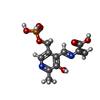  Mass: 316.204 Da / Num. of mol.: 2 / Source method: obtained synthetically / Formula: C11H13N2O7P / Feature type: SUBJECT OF INVESTIGATION Mass: 316.204 Da / Num. of mol.: 2 / Source method: obtained synthetically / Formula: C11H13N2O7P / Feature type: SUBJECT OF INVESTIGATION#4: Water | ChemComp-HOH / |  Mass: 18.015 Da / Num. of mol.: 621 / Source method: isolated from a natural source / Formula: H2O Mass: 18.015 Da / Num. of mol.: 621 / Source method: isolated from a natural source / Formula: H2OHas ligand of interest | Y | Has protein modification | N | |
|---|
-Experimental details
-Experiment
| Experiment | Method: X-RAY DIFFRACTION / Number of used crystals: 1 |
|---|
- Sample preparation
| Crystal | Density Matthews: 2.33 Å3/Da / Density % sol: 47.25 % |
|---|---|
| Crystal grow | Temperature: 291.15 K / Method: vapor diffusion, sitting drop Details: 0.1 M MES, 6.25 % w/v PEG 2000, 6.25 % w/v PEG 3350, 6.25 % w/v PEG 4000, 6.25 % w/v PEG 5000 MME, pH 6.5 |
-Data collection
| Diffraction | Mean temperature: 100 K / Serial crystal experiment: N |
|---|---|
| Diffraction source | Source: SYNCHROTRON / Site: SSRF / Beamline: BL10U2 / Wavelength: 0.97918 Å |
| Detector | Type: DECTRIS EIGER X 16M / Detector: PIXEL / Date: Oct 21, 2024 |
| Radiation | Protocol: SINGLE WAVELENGTH / Monochromatic (M) / Laue (L): M / Scattering type: x-ray |
| Radiation wavelength | Wavelength: 0.97918 Å / Relative weight: 1 |
| Reflection | Resolution: 2.1→38.58 Å / Num. obs: 47945 / % possible obs: 97.5 % / Redundancy: 2.8 % / CC1/2: 0.98 / Rmerge(I) obs: 0.058 / Rpim(I) all: 0.054 / Rrim(I) all: 0.08 / Χ2: 0.52 / Net I/σ(I): 8.8 |
| Reflection shell | Resolution: 2.1→2.16 Å / Redundancy: 1.6 % / Rmerge(I) obs: 0.153 / Num. unique obs: 3524 / CC1/2: 0.932 / Rpim(I) all: 0.152 / Rrim(I) all: 0.216 / Χ2: 0.75 |
- Processing
| Software |
| |||||||||||||||||||||||||||||||||||||||||||||||||||||||||||||||||||||||||||||||||||||||||||||||||||||||||
|---|---|---|---|---|---|---|---|---|---|---|---|---|---|---|---|---|---|---|---|---|---|---|---|---|---|---|---|---|---|---|---|---|---|---|---|---|---|---|---|---|---|---|---|---|---|---|---|---|---|---|---|---|---|---|---|---|---|---|---|---|---|---|---|---|---|---|---|---|---|---|---|---|---|---|---|---|---|---|---|---|---|---|---|---|---|---|---|---|---|---|---|---|---|---|---|---|---|---|---|---|---|---|---|---|---|---|
| Refinement | Method to determine structure: MOLECULAR REPLACEMENT / Resolution: 2.1→36.81 Å / SU ML: 0.18 / Cross valid method: FREE R-VALUE / σ(F): 1.38 / Phase error: 18.4 / Stereochemistry target values: ML
| |||||||||||||||||||||||||||||||||||||||||||||||||||||||||||||||||||||||||||||||||||||||||||||||||||||||||
| Solvent computation | Shrinkage radii: 0.9 Å / VDW probe radii: 1.1 Å / Solvent model: FLAT BULK SOLVENT MODEL | |||||||||||||||||||||||||||||||||||||||||||||||||||||||||||||||||||||||||||||||||||||||||||||||||||||||||
| Refinement step | Cycle: LAST / Resolution: 2.1→36.81 Å
| |||||||||||||||||||||||||||||||||||||||||||||||||||||||||||||||||||||||||||||||||||||||||||||||||||||||||
| Refine LS restraints |
| |||||||||||||||||||||||||||||||||||||||||||||||||||||||||||||||||||||||||||||||||||||||||||||||||||||||||
| LS refinement shell |
|
