 Movie
Movie Controller

[English] 日本語
 Yorodumi
- PDB-9wwe: Crystal structure of human tyrosylprotein sulfotransferase 2 (TPS... -
+ Open data

- Basic information
| Entry | Database: PDB / ID: 9wwe | ||||||||||||
|---|---|---|---|---|---|---|---|---|---|---|---|---|---|
| Title | Crystal structure of human tyrosylprotein sulfotransferase 2 (TPST2) in cation-bound state | ||||||||||||
 Components Components | Protein-tyrosine sulfotransferase 2 | ||||||||||||
 Keywords Keywords | PEPTIDE BINDING PROTEIN / TPST2 / Sulfotransferase / Cation | ||||||||||||
| Function / homology |  Function and homology information Function and homology informationpeptidyl-tyrosine sulfation / protein-tyrosine sulfotransferase / protein-tyrosine sulfotransferase activity / prevention of polyspermy / Defective F8 sulfation at Y1699 / Cytosolic sulfonation of small molecules / Gamma carboxylation, hypusinylation, hydroxylation, and arylsulfatase activation / fusion of sperm to egg plasma membrane involved in single fertilization / 3'-phosphoadenosine 5'-phosphosulfate metabolic process / trans-Golgi network ...peptidyl-tyrosine sulfation / protein-tyrosine sulfotransferase / protein-tyrosine sulfotransferase activity / prevention of polyspermy / Defective F8 sulfation at Y1699 / Cytosolic sulfonation of small molecules / Gamma carboxylation, hypusinylation, hydroxylation, and arylsulfatase activation / fusion of sperm to egg plasma membrane involved in single fertilization / 3'-phosphoadenosine 5'-phosphosulfate metabolic process / trans-Golgi network / Golgi lumen / Golgi membrane / endoplasmic reticulum / Golgi apparatus / protein homodimerization activity Similarity search - Function | ||||||||||||
| Biological species |  Homo sapiens (human) Homo sapiens (human) | ||||||||||||
| Method |  X-RAY DIFFRACTION / SYNCHROTRON / MOLECULAR REPLACEMENT / Resolution: 1.75 Å X-RAY DIFFRACTION / SYNCHROTRON / MOLECULAR REPLACEMENT / Resolution: 1.75 Å | ||||||||||||
 Authors Authors | Jin, M. / Yang, J. / Kim, H. / Eom, S.H. | ||||||||||||
| Funding support |  Korea, Republic Of, 3items Korea, Republic Of, 3items
| ||||||||||||
 Citation Citation | Journal: To Be Published Title: Crystal structure of human tyrosylprotein sulfotransferase 2 (TPST2) in cation-bound state Authors: Jin, M. / Yang, J. / Kim, H. / Eom, S.H. | ||||||||||||
| History |
|
- Structure visualization
| Structure viewer | Molecule: MolmilJmol/JSmol |
|---|
- Downloads & links
-Download
| PDBx/mmCIF format | 9wwe.cif.gz | 101.9 KB | Display | PDBx/mmCIF format |
|---|---|---|---|---|
| PDB format | pdb9wwe.ent.gz | 60 KB | Display | PDB format |
| PDBx/mmJSON format | 9wwe.json.gz | Tree view | PDBx/mmJSON format | |
| Others |  Other downloads Other downloads |
-Validation report
| Arichive directory | https://data.pdbj.org/pub/pdb/validation_reports/ww/9wweftp://data.pdbj.org/pub/pdb/validation_reports/ww/9wwe | HTTPS FTP |
|---|
-Related structure data
| Similar structure data |
|---|
-Links
 PDBj
PDBj

- Assembly
| Deposited unit | 
| ||||||||||
|---|---|---|---|---|---|---|---|---|---|---|---|
| 1 | 
| ||||||||||
| Unit cell |
| ||||||||||
| Components on special symmetry positions |
|
-Components
-Protein , 1 types, 1 molecules A
| #1: Protein | Mass: 35957.715 Da / Num. of mol.: 1 Source method: isolated from a genetically manipulated source Source: (gene. exp.) Homo sapiens (human) / Gene: TPST2 / Production host:  References: UniProt: O60704, protein-tyrosine sulfotransferase |
|---|
-Non-polymers , 6 types, 265 molecules 

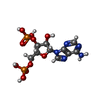


| #2: Chemical |  Mass: 22.990 Da / Num. of mol.: 2 / Source method: obtained synthetically / Formula: Na Mass: 22.990 Da / Num. of mol.: 2 / Source method: obtained synthetically / Formula: Na#3: Chemical |  Mass: 65.409 Da / Num. of mol.: 2 / Source method: obtained synthetically / Formula: Zn Mass: 65.409 Da / Num. of mol.: 2 / Source method: obtained synthetically / Formula: Zn#4: Chemical | ChemComp-A3P / |  Type: RNA linking / Mass: 427.201 Da / Num. of mol.: 1 / Source method: obtained synthetically / Formula: C10H15N5O10P2 Type: RNA linking / Mass: 427.201 Da / Num. of mol.: 1 / Source method: obtained synthetically / Formula: C10H15N5O10P2#5: Chemical | ChemComp-GOL / |  Mass: 92.094 Da / Num. of mol.: 1 / Source method: obtained synthetically / Formula: C3H8O3 Mass: 92.094 Da / Num. of mol.: 1 / Source method: obtained synthetically / Formula: C3H8O3#6: Chemical | ChemComp-TLA / |  Mass: 150.087 Da / Num. of mol.: 1 / Source method: obtained synthetically / Formula: C4H6O6 / Feature type: SUBJECT OF INVESTIGATION Mass: 150.087 Da / Num. of mol.: 1 / Source method: obtained synthetically / Formula: C4H6O6 / Feature type: SUBJECT OF INVESTIGATION#7: Water | ChemComp-HOH / | Mass: 18.015 Da / Num. of mol.: 258 / Source method: isolated from a natural source / Formula: H2O |
|---|
-Details
| Has ligand of interest | Y |
|---|---|
| Has protein modification | Y |
-Experimental details
-Experiment
| Experiment | Method: X-RAY DIFFRACTION / Number of used crystals: 1 |
|---|
- Sample preparation
| Crystal | Density Matthews: 3.77 Å3/Da / Density % sol: 67.34 % |
|---|---|
| Crystal grow | Temperature: 293.15 K / Method: vapor diffusion Details: 0.1M HEPES-NaOH (pH 7.0), 16% (w/v) PEG 6000, 0.1M Potassium sodium tartrate, 20mM ZnCl2 |
-Data collection
| Diffraction | Mean temperature: 80 K / Serial crystal experiment: N |
|---|---|
| Diffraction source | Source: SYNCHROTRON / Site: PAL/PLS / Beamline: 5C (4A) / Wavelength: 1 Å |
| Detector | Type: DECTRIS EIGER2 X 9M / Detector: PIXEL / Date: Apr 2, 2025 |
| Radiation | Protocol: SINGLE WAVELENGTH / Monochromatic (M) / Laue (L): M / Scattering type: x-ray |
| Radiation wavelength | Wavelength: 1 Å / Relative weight: 1 |
| Reflection | Resolution: 1.75→46.21 Å / Num. obs: 55466 / % possible obs: 100 % / Redundancy: 26.8 % / CC1/2: 0.999 / Rmerge(I) obs: 0.144 / Χ2: 1.02 / Net I/σ(I): 18.7 |
| Reflection shell | Resolution: 1.75→1.79 Å / Redundancy: 26.3 % / Rmerge(I) obs: 1.235 / Mean I/σ(I) obs: 3.5 / Num. unique obs: 2964 / CC1/2: 0.76 / Χ2: 0.99 / % possible all: 99.2 |
- Processing
| Software |
| |||||||||||||||||||||||||||||||||||||||||||||||||||||||||||||||||||||||||||||||||||||||||||||||||||||||||
|---|---|---|---|---|---|---|---|---|---|---|---|---|---|---|---|---|---|---|---|---|---|---|---|---|---|---|---|---|---|---|---|---|---|---|---|---|---|---|---|---|---|---|---|---|---|---|---|---|---|---|---|---|---|---|---|---|---|---|---|---|---|---|---|---|---|---|---|---|---|---|---|---|---|---|---|---|---|---|---|---|---|---|---|---|---|---|---|---|---|---|---|---|---|---|---|---|---|---|---|---|---|---|---|---|---|---|
| Refinement | Method to determine structure: MOLECULAR REPLACEMENT / Resolution: 1.75→46.21 Å / SU ML: 0.1501 / Cross valid method: FREE R-VALUE / σ(F): 1.34 / Phase error: 17.0064 Stereochemistry target values: GeoStd + Monomer Library + CDL v1.2
| |||||||||||||||||||||||||||||||||||||||||||||||||||||||||||||||||||||||||||||||||||||||||||||||||||||||||
| Solvent computation | Shrinkage radii: 0.9 Å / VDW probe radii: 1.1 Å / Solvent model: FLAT BULK SOLVENT MODEL | |||||||||||||||||||||||||||||||||||||||||||||||||||||||||||||||||||||||||||||||||||||||||||||||||||||||||
| Displacement parameters | Biso mean: 24.29 Å2 | |||||||||||||||||||||||||||||||||||||||||||||||||||||||||||||||||||||||||||||||||||||||||||||||||||||||||
| Refinement step | Cycle: LAST / Resolution: 1.75→46.21 Å
| |||||||||||||||||||||||||||||||||||||||||||||||||||||||||||||||||||||||||||||||||||||||||||||||||||||||||
| Refine LS restraints |
| |||||||||||||||||||||||||||||||||||||||||||||||||||||||||||||||||||||||||||||||||||||||||||||||||||||||||
| LS refinement shell |
|
