 Movie
Movie Controller

[English] 日本語
 Yorodumi
- PDB-9wp1: Structural insights into tyrosine sulfation of CCR5 by human tyro... -
+ Open data

- Basic information
| Entry | Database: PDB / ID: 9wp1 | ||||||||||||
|---|---|---|---|---|---|---|---|---|---|---|---|---|---|
| Title | Structural insights into tyrosine sulfation of CCR5 by human tyrosylprotein sulfotransferase-1 | ||||||||||||
 Components Components |
| ||||||||||||
 Keywords Keywords | TRANSFERASE / sulfotransferase / sulfation | ||||||||||||
| Function / homology |  Function and homology information Function and homology informationpeptidyl-tyrosine sulfation / protein-tyrosine sulfotransferase / protein-tyrosine sulfotransferase activity / chemokine (C-C motif) ligand 5 binding / Defective F8 sulfation at Y1699 / Cytosolic sulfonation of small molecules / Gamma carboxylation, hypusinylation, hydroxylation, and arylsulfatase activation / 3'-phosphoadenosine 5'-phosphosulfate metabolic process / negative regulation of macrophage apoptotic process / chemokine receptor activity ...peptidyl-tyrosine sulfation / protein-tyrosine sulfotransferase / protein-tyrosine sulfotransferase activity / chemokine (C-C motif) ligand 5 binding / Defective F8 sulfation at Y1699 / Cytosolic sulfonation of small molecules / Gamma carboxylation, hypusinylation, hydroxylation, and arylsulfatase activation / 3'-phosphoadenosine 5'-phosphosulfate metabolic process / negative regulation of macrophage apoptotic process / chemokine receptor activity / signaling / phosphatidylinositol-4,5-bisphosphate phospholipase C activity / C-C chemokine binding / response to cholesterol / C-C chemokine receptor activity / Chemokine receptors bind chemokines / release of sequestered calcium ion into cytosol by sarcoplasmic reticulum / dendritic cell chemotaxis / Interleukin-10 signaling / Binding and entry of HIV virion / cellular defense response / coreceptor activity / post-translational protein modification / calcium-mediated signaling / cell chemotaxis / trans-Golgi network / Golgi lumen / chemotaxis / calcium ion transport / MAPK cascade / cell-cell signaling / cellular response to lipopolysaccharide / positive regulation of cytosolic calcium ion concentration / virus receptor activity / actin binding / G alpha (i) signalling events / cell surface receptor signaling pathway / endosome / immune response / inflammatory response / G protein-coupled receptor signaling pathway / external side of plasma membrane / Golgi membrane / Golgi apparatus / cell surface / protein homodimerization activity / membrane / identical protein binding / plasma membrane / cytoplasm Similarity search - Function | ||||||||||||
| Biological species |  Homo sapiens (human) Homo sapiens (human) | ||||||||||||
| Method |  X-RAY DIFFRACTION / SYNCHROTRON / MOLECULAR REPLACEMENT / Resolution: 3.2 Å X-RAY DIFFRACTION / SYNCHROTRON / MOLECULAR REPLACEMENT / Resolution: 3.2 Å | ||||||||||||
 Authors Authors | Tanaka, S. / Asano, H. / Toyoda, K. / Nishiyori, T. / Kojo, H. / Nishimoto, E. / Teramoto, T. / Kakuta, Y. | ||||||||||||
| Funding support |  Japan, 3items Japan, 3items
| ||||||||||||
 Citation Citation | Journal: Febs J. / Year: 2026 Title: Structural insights into tyrosine sulfation of CCR5 by human tyrosylprotein sulfotransferase-1. Authors: Tanaka, S. / Asano, H. / Toyoda, K. / Nishiyori, T. / Kojo, H. / Kiyomatsu, K. / Kurogi, K. / Sakakibara, Y. / Nishimoto, E. / Teramoto, T. / Kakuta, Y. | ||||||||||||
| History |
|
- Structure visualization
| Structure viewer | Molecule: MolmilJmol/JSmol |
|---|
- Downloads & links
-Download
| PDBx/mmCIF format | 9wp1.cif.gz | 444.9 KB | Display | PDBx/mmCIF format |
|---|---|---|---|---|
| PDB format | pdb9wp1.ent.gz | 366.2 KB | Display | PDB format |
| PDBx/mmJSON format | 9wp1.json.gz | Tree view | PDBx/mmJSON format | |
| Others |  Other downloads Other downloads |
-Validation report
| Arichive directory | https://data.pdbj.org/pub/pdb/validation_reports/wp/9wp1ftp://data.pdbj.org/pub/pdb/validation_reports/wp/9wp1 | HTTPS FTP |
|---|
-Related structure data
| Related structure data |  3ap1S S: Starting model for refinement |
|---|---|
| Similar structure data |


-Links
 PDBj
PDBj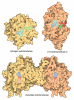








- Assembly
| Deposited unit | 
| ||||||||
|---|---|---|---|---|---|---|---|---|---|
| 1 |
| ||||||||
| 2 | 
| ||||||||
| 3 | 
| ||||||||
| 4 | 
| ||||||||
| 5 | 
| ||||||||
| Unit cell |
|
-Components
| #1: Protein | Mass: 31366.777 Da / Num. of mol.: 8 Source method: isolated from a genetically manipulated source Source: (gene. exp.) Homo sapiens (human) / Gene: TPST1 / Production host:   Escherichia coli (E. coli) Escherichia coli (E. coli)References: UniProt: O60507, protein-tyrosine sulfotransferase #2: Protein/peptide | Mass: 741.810 Da / Num. of mol.: 8 / Source method: obtained synthetically / Source: (synth.) Homo sapiens (human) / References: UniProt: P51681#3: Chemical | ChemComp-A3P / 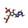 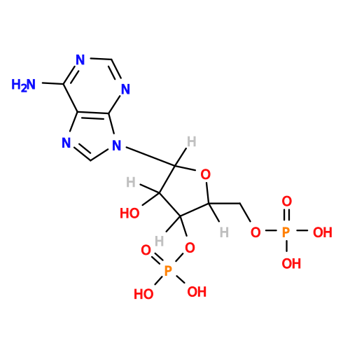 Type: RNA linking / Mass: 427.201 Da / Num. of mol.: 8 / Source method: obtained synthetically / Formula: C10H15N5O10P2 / Feature type: SUBJECT OF INVESTIGATION Type: RNA linking / Mass: 427.201 Da / Num. of mol.: 8 / Source method: obtained synthetically / Formula: C10H15N5O10P2 / Feature type: SUBJECT OF INVESTIGATION#4: Chemical | ChemComp-MG /   Mass: 24.305 Da / Num. of mol.: 8 / Source method: obtained synthetically / Formula: Mg / Feature type: SUBJECT OF INVESTIGATION Mass: 24.305 Da / Num. of mol.: 8 / Source method: obtained synthetically / Formula: Mg / Feature type: SUBJECT OF INVESTIGATION#5: Chemical | ChemComp-ZN /   Mass: 65.409 Da / Num. of mol.: 4 / Source method: obtained synthetically / Formula: Zn / Feature type: SUBJECT OF INVESTIGATION Mass: 65.409 Da / Num. of mol.: 4 / Source method: obtained synthetically / Formula: Zn / Feature type: SUBJECT OF INVESTIGATIONHas ligand of interest | Y | Has protein modification | Y | |
|---|
-Experimental details
-Experiment
| Experiment | Method: X-RAY DIFFRACTION / Number of used crystals: 1 |
|---|
- Sample preparation
| Crystal | Density Matthews: 2.88 Å3/Da / Density % sol: 57.24 % |
|---|---|
| Crystal grow | Temperature: 293 K / Method: vapor diffusion, hanging drop / pH: 6.9 / Details: 1.8 M sodium/potassium phosphate, |
-Data collection
| Diffraction | Mean temperature: 100 K / Serial crystal experiment: N |
|---|---|
| Diffraction source | Source: SYNCHROTRON / Site: SPring-8 / Beamline: BL26B2 / Wavelength: 1 Å |
| Detector | Type: MARMOSAIC 225 mm CCD / Detector: CCD / Date: Sep 1, 2020 |
| Radiation | Protocol: SINGLE WAVELENGTH / Monochromatic (M) / Laue (L): M / Scattering type: x-ray |
| Radiation wavelength | Wavelength: 1 Å / Relative weight: 1 |
| Reflection | Resolution: 3.2→50 Å / Num. obs: 56568 / % possible obs: 99.9 % / Redundancy: 3.7 % / CC1/2: 0.99 / Net I/σ(I): 2.73 |
| Reflection shell | Resolution: 3.2→3.26 Å / Num. unique obs: 5656 / CC1/2: 0.484 |
- Processing
| Software |
| ||||||||||||||||||||||||||||||||||||||||||||||||||||||||||||||||||||||||||||||||||||||||||||||||||||||||||||||||||||||||||||||||||||||||||||||||||||||||||
|---|---|---|---|---|---|---|---|---|---|---|---|---|---|---|---|---|---|---|---|---|---|---|---|---|---|---|---|---|---|---|---|---|---|---|---|---|---|---|---|---|---|---|---|---|---|---|---|---|---|---|---|---|---|---|---|---|---|---|---|---|---|---|---|---|---|---|---|---|---|---|---|---|---|---|---|---|---|---|---|---|---|---|---|---|---|---|---|---|---|---|---|---|---|---|---|---|---|---|---|---|---|---|---|---|---|---|---|---|---|---|---|---|---|---|---|---|---|---|---|---|---|---|---|---|---|---|---|---|---|---|---|---|---|---|---|---|---|---|---|---|---|---|---|---|---|---|---|---|---|---|---|---|---|---|---|
| Refinement | Method to determine structure: MOLECULAR REPLACEMENT Starting model: 3AP1 Resolution: 3.2→49.21 Å / SU ML: 0.55 / Cross valid method: FREE R-VALUE / σ(F): 1.35 / Phase error: 34.61 / Stereochemistry target values: ML
| ||||||||||||||||||||||||||||||||||||||||||||||||||||||||||||||||||||||||||||||||||||||||||||||||||||||||||||||||||||||||||||||||||||||||||||||||||||||||||
| Solvent computation | Shrinkage radii: 0.9 Å / VDW probe radii: 1.1 Å / Solvent model: FLAT BULK SOLVENT MODEL | ||||||||||||||||||||||||||||||||||||||||||||||||||||||||||||||||||||||||||||||||||||||||||||||||||||||||||||||||||||||||||||||||||||||||||||||||||||||||||
| Refinement step | Cycle: LAST / Resolution: 3.2→49.21 Å
| ||||||||||||||||||||||||||||||||||||||||||||||||||||||||||||||||||||||||||||||||||||||||||||||||||||||||||||||||||||||||||||||||||||||||||||||||||||||||||
| Refine LS restraints |
| ||||||||||||||||||||||||||||||||||||||||||||||||||||||||||||||||||||||||||||||||||||||||||||||||||||||||||||||||||||||||||||||||||||||||||||||||||||||||||
| LS refinement shell |
|
