 Movie
Movie Controller

[English] 日本語
 Yorodumi
- PDB-9weg: Plasmodium vivax aspartyl-tRNA synthetase in complex with AMS, Mg... -
+ Open data

- Basic information
| Entry | Database: PDB / ID: 9weg | |||||||||
|---|---|---|---|---|---|---|---|---|---|---|
| Title | Plasmodium vivax aspartyl-tRNA synthetase in complex with AMS, Mg ion, Iodides and Pentaethylene glycol | |||||||||
 Components Components | aspartate--tRNA ligase | |||||||||
 Keywords Keywords | LIGASE / AMINOACYLATION / AMINOACYL-TRNA SYNTHETASE / TRNA-BINDING / ATP-BINDING / MALARIA / INHIBITOR | |||||||||
| Function / homology |  Function and homology information Function and homology informationaspartate-tRNA ligase / aspartate-tRNA ligase activity / aspartyl-tRNA aminoacylation / aminoacyl-tRNA synthetase multienzyme complex / RNA binding / ATP binding / cytosol Similarity search - Function | |||||||||
| Biological species |   Plasmodium vivax (malaria parasite P. vivax) Plasmodium vivax (malaria parasite P. vivax) | |||||||||
| Method |  X-RAY DIFFRACTION / SYNCHROTRON / MOLECULAR REPLACEMENT / Resolution: 2.228 Å X-RAY DIFFRACTION / SYNCHROTRON / MOLECULAR REPLACEMENT / Resolution: 2.228 Å | |||||||||
 Authors Authors | Manickam, Y. / Sharma, V.K. / Sharma, A. | |||||||||
| Funding support |  India, 2items India, 2items
| |||||||||
 Citation Citation | Journal: To Be Published Title: The active site of aspartyl-tRNA synthetase: Structural studies of the adenylation reaction and flexibility of residues. Authors: Sharma, V.K. / Manickam, Y. / Sharma, A. | |||||||||
| History |
|
- Structure visualization
| Structure viewer | Molecule: MolmilJmol/JSmol |
|---|
- Downloads & links
-Download
| PDBx/mmCIF format | 9weg.cif.gz | 447.5 KB | Display | PDBx/mmCIF format |
|---|---|---|---|---|
| PDB format | pdb9weg.ent.gz | 363 KB | Display | PDB format |
| PDBx/mmJSON format | 9weg.json.gz | Tree view | PDBx/mmJSON format | |
| Others |  Other downloads Other downloads |
-Validation report
| Arichive directory | https://data.pdbj.org/pub/pdb/validation_reports/we/9wegftp://data.pdbj.org/pub/pdb/validation_reports/we/9weg | HTTPS FTP |
|---|
-Related structure data
| Related structure data |  9we8C  9we9C  9weaC  9webC  9wecC  9wedC  9weeC  9wefC  9wehC  9weiC  9wejC  9wekC  9welC  9wemC C: citing same article ( |
|---|---|
| Similar structure data |
-Links
 PDBj
PDBj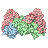
- Assembly
| Deposited unit | 
| ||||||||
|---|---|---|---|---|---|---|---|---|---|
| 1 |
| ||||||||
| Unit cell |
| ||||||||
| Components on special symmetry positions |
|
-Components
-Protein , 1 types, 2 molecules AB
| #1: Protein | Mass: 62128.812 Da / Num. of mol.: 2 Source method: isolated from a genetically manipulated source Source: (gene. exp.) Plasmodium vivax (malaria parasite P. vivax)Gene: PVC01_020016700, PVW1_020019400 / Production host:   Escherichia coli (E. coli) / References: UniProt: A0A1G4H6Y1, aspartate-tRNA ligase Escherichia coli (E. coli) / References: UniProt: A0A1G4H6Y1, aspartate-tRNA ligase |
|---|
-Non-polymers , 6 types, 385 molecules 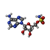


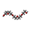

| #2: Chemical | 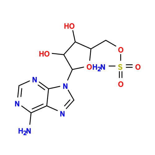 Type: RNA linking / Mass: 346.320 Da / Num. of mol.: 2 / Source method: obtained synthetically / Formula: C10H14N6O6S / Feature type: SUBJECT OF INVESTIGATION Type: RNA linking / Mass: 346.320 Da / Num. of mol.: 2 / Source method: obtained synthetically / Formula: C10H14N6O6S / Feature type: SUBJECT OF INVESTIGATION#3: Chemical | ChemComp-IOD /  Mass: 126.904 Da / Num. of mol.: 5 / Source method: obtained synthetically / Formula: I Mass: 126.904 Da / Num. of mol.: 5 / Source method: obtained synthetically / Formula: I#4: Chemical | ChemComp-GOL / | 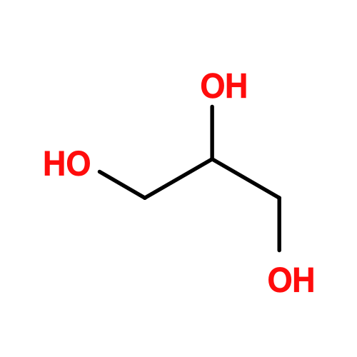 Mass: 92.094 Da / Num. of mol.: 1 / Source method: obtained synthetically / Formula: C3H8O3 Mass: 92.094 Da / Num. of mol.: 1 / Source method: obtained synthetically / Formula: C3H8O3#5: Chemical | ChemComp-MG / |  Mass: 24.305 Da / Num. of mol.: 1 / Source method: obtained synthetically / Formula: Mg Mass: 24.305 Da / Num. of mol.: 1 / Source method: obtained synthetically / Formula: Mg#6: Chemical | ChemComp-1PE / | 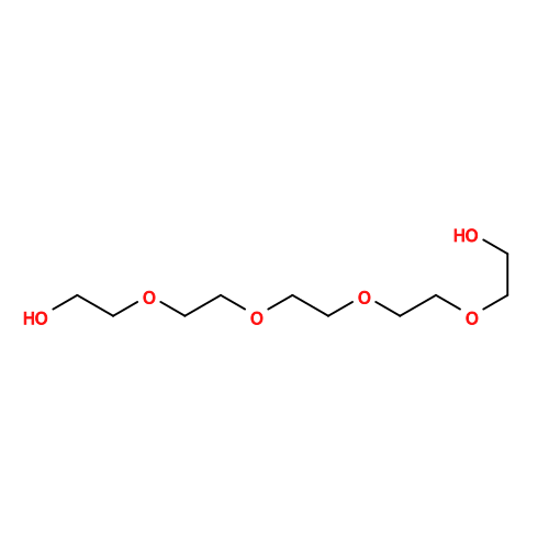 Mass: 238.278 Da / Num. of mol.: 1 / Source method: obtained synthetically / Formula: C10H22O6 / Comment: precipitant*YM Mass: 238.278 Da / Num. of mol.: 1 / Source method: obtained synthetically / Formula: C10H22O6 / Comment: precipitant*YM#7: Water | ChemComp-HOH / | Mass: 18.015 Da / Num. of mol.: 375 / Source method: isolated from a natural source / Formula: H2O |
|---|
-Details
| Has ligand of interest | Y |
|---|---|
| Has protein modification | N |
-Experimental details
-Experiment
| Experiment | Method: X-RAY DIFFRACTION / Number of used crystals: 1 |
|---|
- Sample preparation
| Crystal | Density Matthews: 3.07 Å3/Da / Density % sol: 59.96 % |
|---|---|
| Crystal grow | Temperature: 293 K / Method: vapor diffusion, hanging drop Details: Morpheus B5: 0.1 M Buffer System 2 pH 7.5 (Sodium HEPES and MOPS), 30% Precipitant Mix 1 (40% v/v PEG 500* MME; 20 % w/v PEG 20000) and 0.09 M Halogens (0.3 M Sodium fluoride; 0.3 M Sodium ...Details: Morpheus B5: 0.1 M Buffer System 2 pH 7.5 (Sodium HEPES and MOPS), 30% Precipitant Mix 1 (40% v/v PEG 500* MME; 20 % w/v PEG 20000) and 0.09 M Halogens (0.3 M Sodium fluoride; 0.3 M Sodium bromide; 0.3 M Sodium iodide) |
-Data collection
| Diffraction | Mean temperature: 100 K / Serial crystal experiment: N |
|---|---|
| Diffraction source | Source: SYNCHROTRON / Site: Diamond  / Beamline: I03 / Wavelength: 0.97627 Å / Beamline: I03 / Wavelength: 0.97627 Å |
| Detector | Type: DECTRIS EIGER X 16M / Detector: PIXEL / Date: Nov 26, 2022 |
| Radiation | Protocol: SINGLE WAVELENGTH / Monochromatic (M) / Laue (L): M / Scattering type: x-ray |
| Radiation wavelength | Wavelength: 0.97627 Å / Relative weight: 1 |
| Reflection | Resolution: 2.228→120.105 Å / Num. obs: 76798 / % possible obs: 100 % / Redundancy: 37.6 % / CC1/2: 0.997 / Rrim(I) all: 0.196 / Net I/σ(I): 16.2 |
| Reflection shell | Resolution: 2.228→2.267 Å / Redundancy: 20.9 % / Mean I/σ(I) obs: 0.5 / Num. unique obs: 3784 / CC1/2: 0.261 / % possible all: 100 |
- Processing
| Software |
| |||||||||||||||||||||||||||||||||||||||||||||||||||||||||||||||||||||||||||||||||||||||||||||||||||||||||||||||||||||||||||||||||||||||||||||||||||||||||||||||||||||||||||||||||||||||||||||||||||||||||||
|---|---|---|---|---|---|---|---|---|---|---|---|---|---|---|---|---|---|---|---|---|---|---|---|---|---|---|---|---|---|---|---|---|---|---|---|---|---|---|---|---|---|---|---|---|---|---|---|---|---|---|---|---|---|---|---|---|---|---|---|---|---|---|---|---|---|---|---|---|---|---|---|---|---|---|---|---|---|---|---|---|---|---|---|---|---|---|---|---|---|---|---|---|---|---|---|---|---|---|---|---|---|---|---|---|---|---|---|---|---|---|---|---|---|---|---|---|---|---|---|---|---|---|---|---|---|---|---|---|---|---|---|---|---|---|---|---|---|---|---|---|---|---|---|---|---|---|---|---|---|---|---|---|---|---|---|---|---|---|---|---|---|---|---|---|---|---|---|---|---|---|---|---|---|---|---|---|---|---|---|---|---|---|---|---|---|---|---|---|---|---|---|---|---|---|---|---|---|---|---|---|---|---|---|---|
| Refinement | Method to determine structure: MOLECULAR REPLACEMENT / Resolution: 2.228→45.832 Å / SU ML: 0.27 / Cross valid method: FREE R-VALUE / σ(F): 1.33 / Phase error: 23.47 / Stereochemistry target values: ML
| |||||||||||||||||||||||||||||||||||||||||||||||||||||||||||||||||||||||||||||||||||||||||||||||||||||||||||||||||||||||||||||||||||||||||||||||||||||||||||||||||||||||||||||||||||||||||||||||||||||||||||
| Solvent computation | Shrinkage radii: 0.9 Å / VDW probe radii: 1.11 Å / Solvent model: FLAT BULK SOLVENT MODEL | |||||||||||||||||||||||||||||||||||||||||||||||||||||||||||||||||||||||||||||||||||||||||||||||||||||||||||||||||||||||||||||||||||||||||||||||||||||||||||||||||||||||||||||||||||||||||||||||||||||||||||
| Refinement step | Cycle: LAST / Resolution: 2.228→45.832 Å
| |||||||||||||||||||||||||||||||||||||||||||||||||||||||||||||||||||||||||||||||||||||||||||||||||||||||||||||||||||||||||||||||||||||||||||||||||||||||||||||||||||||||||||||||||||||||||||||||||||||||||||
| Refine LS restraints |
| |||||||||||||||||||||||||||||||||||||||||||||||||||||||||||||||||||||||||||||||||||||||||||||||||||||||||||||||||||||||||||||||||||||||||||||||||||||||||||||||||||||||||||||||||||||||||||||||||||||||||||
| LS refinement shell |
| |||||||||||||||||||||||||||||||||||||||||||||||||||||||||||||||||||||||||||||||||||||||||||||||||||||||||||||||||||||||||||||||||||||||||||||||||||||||||||||||||||||||||||||||||||||||||||||||||||||||||||
| Refinement TLS params. | Method: refined / Origin x: 18.1619 Å / Origin y: 54.5191 Å / Origin z: 14.1348 Å
| |||||||||||||||||||||||||||||||||||||||||||||||||||||||||||||||||||||||||||||||||||||||||||||||||||||||||||||||||||||||||||||||||||||||||||||||||||||||||||||||||||||||||||||||||||||||||||||||||||||||||||
| Refinement TLS group | Selection details: all |
