 Movie
Movie Controller


+ Open data

- Basic information
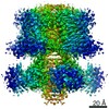
| Entry | Database: PDB / ID: 9wd8 | ||||||
|---|---|---|---|---|---|---|---|
| Title | structure of human KCNQ1-KCNE3-CaM complex with two PIP2 | ||||||
 Components Components |
| ||||||
 Keywords Keywords | MEMBRANE PROTEIN / potassium channel complex | ||||||
| Function / homology |  Function and homology information Function and homology informationnegative regulation of membrane repolarization during ventricular cardiac muscle cell action potential / gastrin-induced gastric acid secretion / corticosterone secretion / voltage-gated potassium channel activity involved in atrial cardiac muscle cell action potential repolarization / negative regulation of voltage-gated potassium channel activity / basolateral part of cell / lumenal side of membrane / negative regulation of delayed rectifier potassium channel activity / rhythmic behavior / stomach development ...negative regulation of membrane repolarization during ventricular cardiac muscle cell action potential / gastrin-induced gastric acid secretion / corticosterone secretion / voltage-gated potassium channel activity involved in atrial cardiac muscle cell action potential repolarization / negative regulation of voltage-gated potassium channel activity / basolateral part of cell / lumenal side of membrane / negative regulation of delayed rectifier potassium channel activity / rhythmic behavior / stomach development / regulation of gastric acid secretion / voltage-gated potassium channel activity involved in cardiac muscle cell action potential repolarization / Phase 3 - rapid repolarisation / membrane repolarization during action potential / negative regulation of potassium ion export across plasma membrane / membrane repolarization during atrial cardiac muscle cell action potential / Phase 2 - plateau phase / iodide transport / regulation of atrial cardiac muscle cell membrane repolarization / membrane repolarization during ventricular cardiac muscle cell action potential / intracellular chloride ion homeostasis / potassium ion export across plasma membrane / membrane repolarization during cardiac muscle cell action potential / voltage-gated potassium channel activity involved in ventricular cardiac muscle cell action potential repolarization / atrial cardiac muscle cell action potential / regulation of membrane repolarization / renal sodium ion absorption / auditory receptor cell development / protein phosphatase 1 binding / detection of mechanical stimulus involved in sensory perception of sound / delayed rectifier potassium channel activity / ventricular cardiac muscle cell action potential / potassium ion homeostasis / regulation of ventricular cardiac muscle cell membrane repolarization / positive regulation of potassium ion transmembrane transport / Voltage gated Potassium channels / non-motile cilium assembly / outward rectifier potassium channel activity / intestinal absorption / CaM pathway / Cam-PDE 1 activation / Sodium/Calcium exchangers / inner ear morphogenesis / Calmodulin induced events / cardiac muscle cell contraction / Reduction of cytosolic Ca++ levels / Activation of Ca-permeable Kainate Receptor / CREB1 phosphorylation through the activation of CaMKII/CaMKK/CaMKIV cascasde / Loss of phosphorylation of MECP2 at T308 / CREB1 phosphorylation through the activation of Adenylate Cyclase / adrenergic receptor signaling pathway / negative regulation of high voltage-gated calcium channel activity / PKA activation / CaMK IV-mediated phosphorylation of CREB / Glycogen breakdown (glycogenolysis) / CLEC7A (Dectin-1) induces NFAT activation / renal absorption / negative regulation of ryanodine-sensitive calcium-release channel activity / organelle localization by membrane tethering / Activation of RAC1 downstream of NMDARs / : / autophagosome membrane docking / protein kinase A regulatory subunit binding / ciliary base / protein kinase A catalytic subunit binding / negative regulation of calcium ion export across plasma membrane / regulation of ryanodine-sensitive calcium-release channel activity / regulation of cardiac muscle cell action potential / regulation of heart contraction / neuronal cell body membrane / presynaptic endocytosis / inner ear development / sodium ion transport / Synthesis of IP3 and IP4 in the cytosol / Phase 0 - rapid depolarisation / potassium ion import across plasma membrane / Negative regulation of NMDA receptor-mediated neuronal transmission / Unblocking of NMDA receptors, glutamate binding and activation / RHO GTPases activate PAKs / calcineurin-mediated signaling / regulation of heart rate by cardiac conduction / regulation of cell communication by electrical coupling involved in cardiac conduction / monoatomic ion channel complex / Ion transport by P-type ATPases / Uptake and function of anthrax toxins / cochlea development / protein phosphatase activator activity / Long-term potentiation / Calcineurin activates NFAT / action potential / Regulation of MECP2 expression and activity / DARPP-32 events / potassium channel regulator activity / Smooth Muscle Contraction / voltage-gated potassium channel activity / detection of calcium ion / social behavior / regulation of cardiac muscle contraction / catalytic complex / positive regulation of heart rate Similarity search - Function | ||||||
| Biological species |  Homo sapiens (human) Homo sapiens (human) | ||||||
| Method | ELECTRON MICROSCOPY / single particle reconstruction / cryo EM / Resolution: 3.9 Å | ||||||
 Authors Authors | Cui, C. / Sun, J. | ||||||
| Funding support |  United States, 1items United States, 1items
| ||||||
 Citation Citation | Journal: Cell Res / Year: 2025 Title: Mechanisms of KCNQ1 gating modulation by KCNE1/3 for cell-specific function. Authors: Chenxi Cui / Lu Zhao / Ali A Kermani / Shuzong Du / Tanadet Pipatpolkai / Meiqin Jiang / Sagar Chittori / Yong Zi Tan / Jingyi Shi / Lucie Delemotte / Jianmin Cui / Ji Sun /   Abstract: KCNQ1 potassium channels are essential for physiological processes such as cardiac rhythm and intestinal chloride secretion. KCNE family subunits (KCNE1-5) associate with KCNQ1, conferring distinct ...KCNQ1 potassium channels are essential for physiological processes such as cardiac rhythm and intestinal chloride secretion. KCNE family subunits (KCNE1-5) associate with KCNQ1, conferring distinct properties across various tissues. KCNQ1 activation requires membrane depolarization and phosphatidylinositol 4,5-bisphosphate (PIP2) whose cellular levels are controlled by Gαq-coupled GPCR activation. While modulation of KCNQ1's voltage-dependent activation by KCNE1/3 is well-characterized, their effects on PIP2-dependent gating of KCNQ1 via GPCR signaling remain less understood. Here we resolved structures of KCNQ1-KCNE1 and reassessed the reported KCNQ1-KCNE3 structures with and without PIP2. We revealed that KCNQ1-KCNE1/3 complexes feature two PIP2-binding sites, with KCNE1/3 contributing to a previously overlooked, uncharacterized site involving residues critical for coupling voltage sensor and pore domains. Via this site, KCNE1 and KCNE3 distinctly modulate the PIP2-dependent gating, in addition to the voltage sensitivity, of KCNQ1. Consequently, KCNE3 converts KCNQ1 into a voltage-insensitive PIP2-gated channel governed by GPCR signaling to maintain ion homeostasis in non-excitable cells. KCNE1, by significantly enhancing KCNQ1's PIP2 affinity and resistance to GPCR regulation, forms predominantly voltage-gated channels with KCNQ1 for conducting the slow-delayed rectifier current in excitable cardiac cells. Our study highlights how KCNE1/3 modulates KCNQ1 gating in different cellular contexts, providing insights into tissue-specifically targeting multi-functional channels. | ||||||
| History |
|
- Structure visualization
| Structure viewer | Molecule: MolmilJmol/JSmol |
|---|
- Downloads & links
-Download
| PDBx/mmCIF format | 9wd8.cif.gz | 370.4 KB | Display | PDBx/mmCIF format |
|---|---|---|---|---|
| PDB format | pdb9wd8.ent.gz | Display | PDB format | |
| PDBx/mmJSON format | 9wd8.json.gz | Tree view | PDBx/mmJSON format | |
| Others |  Other downloads Other downloads |
-Validation report
| Arichive directory | https://data.pdbj.org/pub/pdb/validation_reports/wd/9wd8ftp://data.pdbj.org/pub/pdb/validation_reports/wd/9wd8 | HTTPS FTP |
|---|
-Related structure data
| Related structure data |  9vecC  9veiC  9venC  9veoC M: map data used to model this data C: citing same article ( |
|---|---|
| Similar structure data |


-Links
 PDBj
PDBj


- Assembly
| Deposited unit | 
|
|---|---|
| 1 |
|
-Components
| #1: Protein | Mass: 16852.545 Da / Num. of mol.: 4 Source method: isolated from a genetically manipulated source Source: (gene. exp.) Homo sapiens (human) / Gene: CALM1, CALM, CAM, CAM1 / Production host: Homo sapiens (human) / References: UniProt: P0DP23#2: Protein | Mass: 12013.658 Da / Num. of mol.: 4 Source method: isolated from a genetically manipulated source Source: (gene. exp.) Homo sapiens (human) / Gene: KCNE3 / Production host: Homo sapiens (human) / References: UniProt: Q9Y6H6#3: Protein | Mass: 62086.277 Da / Num. of mol.: 4 Source method: isolated from a genetically manipulated source Source: (gene. exp.) Homo sapiens (human) / Gene: KCNQ1, KCNA8, KCNA9, KVLQT1 / Production host: Homo sapiens (human) / References: UniProt: P51787#4: Chemical | ChemComp-CA /   Mass: 40.078 Da / Num. of mol.: 8 / Source method: obtained synthetically / Formula: Ca / Feature type: SUBJECT OF INVESTIGATION Mass: 40.078 Da / Num. of mol.: 8 / Source method: obtained synthetically / Formula: Ca / Feature type: SUBJECT OF INVESTIGATION#5: Chemical | ChemComp-A1BBG / ( Mass: 1023.066 Da / Num. of mol.: 8 / Source method: obtained synthetically / Formula: C45H85O19P3 / Feature type: SUBJECT OF INVESTIGATION Has ligand of interest | Y | Has protein modification | N | |
|---|
-Experimental details
-Experiment
| Experiment | Method: ELECTRON MICROSCOPY |
|---|---|
| EM experiment | Aggregation state: PARTICLE / 3D reconstruction method: single particle reconstruction |
- Sample preparation
| Component | Name: structure of human KCNQ1-KCNE3-CaM complex with two PIP2 Type: COMPLEX / Entity ID: #1-#3 / Source: RECOMBINANT |
|---|---|
| Molecular weight | Experimental value: NO |
| Source (natural) | Organism: Homo sapiens (human) |
| Source (recombinant) | Organism: Homo sapiens (human) |
| Buffer solution | pH: 7.4 |
| Specimen | Embedding applied: NO / Shadowing applied: NO / Staining applied: NO / Vitrification applied: YES |
| Vitrification | Cryogen name: ETHANE |
- Electron microscopy imaging
| Experimental equipment |  Model: Titan Krios / Image courtesy: FEI Company |
|---|---|
| Microscopy | Model: TFS KRIOS |
| Electron gun | Electron source:  FIELD EMISSION GUN / Accelerating voltage: 300 kV / Illumination mode: FLOOD BEAM FIELD EMISSION GUN / Accelerating voltage: 300 kV / Illumination mode: FLOOD BEAM |
| Electron lens | Mode: BRIGHT FIELD / Nominal defocus max: 2400 nm / Nominal defocus min: 1100 nm |
| Image recording | Electron dose: 94 e/Å2 / Film or detector model: GATAN K2 SUMMIT (4k x 4k) |
- Processing
| EM software |
| ||||||||||||
|---|---|---|---|---|---|---|---|---|---|---|---|---|---|
| CTF correction | Type: PHASE FLIPPING AND AMPLITUDE CORRECTION | ||||||||||||
| 3D reconstruction | Resolution: 3.9 Å / Resolution method: FSC 0.143 CUT-OFF / Num. of particles: 88496 / Symmetry type: POINT |
