 Movie
Movie Controller

[English] 日本語
 Yorodumi
- PDB-9wbu: Crystal structure of Mtb cyclic dinucleotide phosphodiesterase by... -
+ Open data

- Basic information
| Entry | Database: PDB / ID: 9wbu | ||||||
|---|---|---|---|---|---|---|---|
| Title | Crystal structure of Mtb cyclic dinucleotide phosphodiesterase by sulfur-modified cyclic dinucleotide analogue | ||||||
 Components Components | Bifunctional oligoribonuclease and PAP phosphatase NrnA | ||||||
 Keywords Keywords | HYDROLASE / Bifunctional oligoribonuclease and PAP phosphatase NrnA | ||||||
| Function / homology |  Function and homology information Function and homology information3'(2'),5'-bisphosphate nucleotidase / 3'(2'),5'-bisphosphate nucleotidase activity / exonuclease activity / Hydrolases; Acting on ester bonds / nucleic acid binding Similarity search - Function | ||||||
| Biological species |  Mycobacterium tuberculosis H37Rv (bacteria) Mycobacterium tuberculosis H37Rv (bacteria) | ||||||
| Method |  X-RAY DIFFRACTION / MOLECULAR REPLACEMENT / Resolution: 2.96 Å X-RAY DIFFRACTION / MOLECULAR REPLACEMENT / Resolution: 2.96 Å | ||||||
 Authors Authors | Hanuman, S.D. / Rajakumara, E. | ||||||
| Funding support |  India, 1items India, 1items
| ||||||
 Citation Citation | Journal: Rsc Chem Biol / Year: 2026 Title: Structural and biochemical insights into the inhibition of Mycobacterium tuberculosis cyclic dinucleotide phosphodiesterase by a sulfur-modified cyclic dinucleotide analog. Authors: Hanuman, D.S. / Neeharika, S. / Murari, S.K. / Yeboah, S.K. / Sintim, H.O. / Rajakumara, E. | ||||||
| History |
|
- Structure visualization
| Structure viewer | Molecule: MolmilJmol/JSmol |
|---|
- Downloads & links
-Download
| PDBx/mmCIF format | 9wbu.cif.gz | 185.6 KB | Display | PDBx/mmCIF format |
|---|---|---|---|---|
| PDB format | pdb9wbu.ent.gz | Display | PDB format | |
| PDBx/mmJSON format | 9wbu.json.gz | Tree view | PDBx/mmJSON format | |
| Others |  Other downloads Other downloads |
-Validation report
| Arichive directory | https://data.pdbj.org/pub/pdb/validation_reports/wb/9wbuftp://data.pdbj.org/pub/pdb/validation_reports/wb/9wbu | HTTPS FTP |
|---|
-Related structure data
| Similar structure data |
|---|
-Links
 PDBj
PDBj- Assembly
| Deposited unit | 
| ||||||||
|---|---|---|---|---|---|---|---|---|---|
| 1 | 
| ||||||||
| 2 | 
| ||||||||
| Unit cell |
|
-Components
| #1: Protein | Mass: 35456.234 Da / Num. of mol.: 3 / Mutation: NO Source method: isolated from a genetically manipulated source Details: Recombinantly purified c-di-NMP Phosphodiesterase (CdnP) of Mycobacterium tuberculosis Source: (gene. exp.) Mycobacterium tuberculosis H37Rv (bacteria)Gene: nrnA, Rv2837c / Plasmid: pET28c / Production host:  Escherichia coli BL21(DE3) (bacteria) Escherichia coli BL21(DE3) (bacteria)References: UniProt: P71615, Hydrolases; Acting on ester bonds, 3'(2'),5'-bisphosphate nucleotidase #2: Chemical | Mass: 674.477 Da / Num. of mol.: 3 / Source method: obtained synthetically / Formula: C20H24N10O11P2S / Feature type: SUBJECT OF INVESTIGATION #3: Chemical | ChemComp-EDO / | 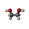 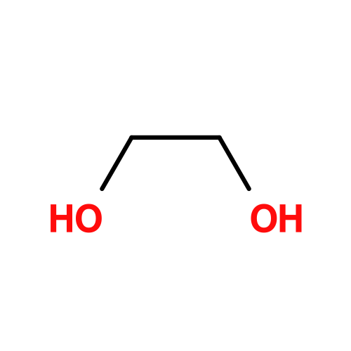 Mass: 62.068 Da / Num. of mol.: 1 / Source method: obtained synthetically / Formula: C2H6O2 Mass: 62.068 Da / Num. of mol.: 1 / Source method: obtained synthetically / Formula: C2H6O2#4: Chemical |   Mass: 54.938 Da / Num. of mol.: 3 / Source method: obtained synthetically / Formula: Mn Mass: 54.938 Da / Num. of mol.: 3 / Source method: obtained synthetically / Formula: MnHas ligand of interest | Y | Has protein modification | N | |
|---|
-Experimental details
-Experiment
| Experiment | Method: X-RAY DIFFRACTION / Number of used crystals: 1 |
|---|
- Sample preparation
| Crystal | Density Matthews: 2.34 Å3/Da / Density % sol: 47.52 % |
|---|---|
| Crystal grow | Temperature: 298 K / Method: vapor diffusion / pH: 4.5 Details: 0.1 M Sodium Acetate Trihydrate pH 4.5, 2M Sodium Formate (30 % Ethylene Glycol) PH range: 4.5 - 5.5 |
-Data collection
| Diffraction | Mean temperature: 100 K / Serial crystal experiment: N |
|---|---|
| Diffraction source | Source: ROTATING ANODE / Type: RIGAKU MICROMAX-007 HF / Wavelength: 1.54179 Å |
| Detector | Type: RIGAKU RAXIS / Detector: IMAGE PLATE / Date: Feb 27, 2025 |
| Radiation | Protocol: SINGLE WAVELENGTH / Monochromatic (M) / Laue (L): M / Scattering type: x-ray |
| Radiation wavelength | Wavelength: 1.54179 Å / Relative weight: 1 |
| Reflection | Resolution: 2.96→51.63 Å / Num. obs: 20959 / % possible obs: 99.86 % / Redundancy: 5.1 % / Biso Wilson estimate: 49.73 Å2 / CC1/2: 0.965 / CC star: 0.991 / Net I/σ(I): 4.8 |
| Reflection shell | Resolution: 2.96→3.14 Å / Redundancy: 5.1 % / Rmerge(I) obs: 1.29 / Num. unique obs: 3344 / CC1/2: 0.73 / Rpim(I) all: 0.63 / Rrim(I) all: 0.39 / Χ2: 0.37 / % possible all: 99.86 |
- Processing
| Software |
| ||||||||||||||||||||||||||||||||||||||||||||||||||||||||||||||||||||||||||||||||||||||||||||||||||||||||||||||||||||||||||||||||||||||||||||||||||||||||||||||||||||||||||||||||||||||
|---|---|---|---|---|---|---|---|---|---|---|---|---|---|---|---|---|---|---|---|---|---|---|---|---|---|---|---|---|---|---|---|---|---|---|---|---|---|---|---|---|---|---|---|---|---|---|---|---|---|---|---|---|---|---|---|---|---|---|---|---|---|---|---|---|---|---|---|---|---|---|---|---|---|---|---|---|---|---|---|---|---|---|---|---|---|---|---|---|---|---|---|---|---|---|---|---|---|---|---|---|---|---|---|---|---|---|---|---|---|---|---|---|---|---|---|---|---|---|---|---|---|---|---|---|---|---|---|---|---|---|---|---|---|---|---|---|---|---|---|---|---|---|---|---|---|---|---|---|---|---|---|---|---|---|---|---|---|---|---|---|---|---|---|---|---|---|---|---|---|---|---|---|---|---|---|---|---|---|---|---|---|---|---|
| Refinement | Method to determine structure: MOLECULAR REPLACEMENT / Resolution: 2.96→25.23 Å / Cor.coef. Fo:Fc: 0.921 / Cor.coef. Fo:Fc free: 0.87 / SU B: 27.935 / SU ML: 0.478 / Cross valid method: THROUGHOUT / ESU R Free: 0.481 / Stereochemistry target values: MAXIMUM LIKELIHOOD / Details: HYDROGENS HAVE BEEN ADDED IN THE RIDING POSITIONS
| ||||||||||||||||||||||||||||||||||||||||||||||||||||||||||||||||||||||||||||||||||||||||||||||||||||||||||||||||||||||||||||||||||||||||||||||||||||||||||||||||||||||||||||||||||||||
| Solvent computation | Ion probe radii: 0.8 Å / Shrinkage radii: 0.8 Å / VDW probe radii: 1.2 Å / Solvent model: MASK | ||||||||||||||||||||||||||||||||||||||||||||||||||||||||||||||||||||||||||||||||||||||||||||||||||||||||||||||||||||||||||||||||||||||||||||||||||||||||||||||||||||||||||||||||||||||
| Displacement parameters | Biso mean: 46.949 Å2
| ||||||||||||||||||||||||||||||||||||||||||||||||||||||||||||||||||||||||||||||||||||||||||||||||||||||||||||||||||||||||||||||||||||||||||||||||||||||||||||||||||||||||||||||||||||||
| Refinement step | Cycle: 1 / Resolution: 2.96→25.23 Å
| ||||||||||||||||||||||||||||||||||||||||||||||||||||||||||||||||||||||||||||||||||||||||||||||||||||||||||||||||||||||||||||||||||||||||||||||||||||||||||||||||||||||||||||||||||||||
| Refine LS restraints |
|