 Movie
Movie Controller


+ Open data

- Basic information
| Entry | Database: PDB / ID: 9w40 | |||||||||||||||||||||
|---|---|---|---|---|---|---|---|---|---|---|---|---|---|---|---|---|---|---|---|---|---|---|
| Title | Structure of adenosine A2BR with an agonist MRS3997 | |||||||||||||||||||||
 Components Components |
| |||||||||||||||||||||
 Keywords Keywords | MEMBRANE PROTEIN / GPCR / membrane protein. / SIGNALING PROTEIN | |||||||||||||||||||||
| Function / homology |  Function and homology information Function and homology informationAdenosine P1 receptors / G protein-coupled adenosine receptor activity / Surfactant metabolism / activation of adenylate cyclase activity / presynaptic modulation of chemical synaptic transmission / vasodilation / electron transport chain / Schaffer collateral - CA1 synapse / Olfactory Signaling Pathway / Activation of the phototransduction cascade ...Adenosine P1 receptors / G protein-coupled adenosine receptor activity / Surfactant metabolism / activation of adenylate cyclase activity / presynaptic modulation of chemical synaptic transmission / vasodilation / electron transport chain / Schaffer collateral - CA1 synapse / Olfactory Signaling Pathway / Activation of the phototransduction cascade / G protein-coupled acetylcholine receptor signaling pathway / G beta:gamma signalling through PLC beta / Presynaptic function of Kainate receptors / Thromboxane signalling through TP receptor / Activation of G protein gated Potassium channels / Inhibition of voltage gated Ca2+ channels via Gbeta/gamma subunits / G-protein activation / Glucagon signaling in metabolic regulation / G beta:gamma signalling through CDC42 / Prostacyclin signalling through prostacyclin receptor / Synthesis, secretion, and inactivation of Glucagon-like Peptide-1 (GLP-1) / photoreceptor disc membrane / G beta:gamma signalling through BTK / ADP signalling through P2Y purinoceptor 12 / Sensory perception of sweet, bitter, and umami (glutamate) taste / Glucagon-type ligand receptors / Adrenaline,noradrenaline inhibits insulin secretion / Vasopressin regulates renal water homeostasis via Aquaporins / G alpha (z) signalling events / Glucagon-like Peptide-1 (GLP1) regulates insulin secretion / ADP signalling through P2Y purinoceptor 1 / cellular response to catecholamine stimulus / G beta:gamma signalling through PI3Kgamma / ADORA2B mediated anti-inflammatory cytokines production / adenylate cyclase-activating dopamine receptor signaling pathway / heterotrimeric G-protein complex / Cooperation of PDCL (PhLP1) and TRiC/CCT in G-protein beta folding / cellular response to prostaglandin E stimulus / GPER1 signaling / presynapse / G alpha (12/13) signalling events / Inactivation, recovery and regulation of the phototransduction cascade / G-protein beta-subunit binding / extracellular vesicle / Thrombin signalling through proteinase activated receptors (PARs) / adenylate cyclase-activating G protein-coupled receptor signaling pathway / signaling receptor complex adaptor activity / GTPase binding / Ca2+ pathway / High laminar flow shear stress activates signaling by PIEZO1 and PECAM1:CDH5:KDR in endothelial cells / G alpha (i) signalling events / G alpha (s) signalling events / G alpha (q) signalling events / Ras protein signal transduction / electron transfer activity / Extra-nuclear estrogen signaling / periplasmic space / iron ion binding / G protein-coupled receptor signaling pathway / lysosomal membrane / heme binding / GTPase activity / synapse / glutamatergic synapse / protein-containing complex binding / signal transduction / extracellular exosome / membrane / plasma membrane / cytosol Similarity search - Function | |||||||||||||||||||||
| Biological species |  Homo sapiens (human) Homo sapiens (human)  Escherichia coli (E. coli) Escherichia coli (E. coli) | |||||||||||||||||||||
| Method | ELECTRON MICROSCOPY / single particle reconstruction / cryo EM / Resolution: 3.2 Å | |||||||||||||||||||||
 Authors Authors | Weng, Y. / Chang, H. / Chen, Y. / Pavan, M. / Gao, Z.G. / Hua, T. / Jacobson, K. / Song, G.J. | |||||||||||||||||||||
| Funding support |  China, 1items China, 1items
| |||||||||||||||||||||
 Citation Citation | Journal: To Be Published Title: Structure of adenosine A2BR with an agonist MRS3997 Authors: Song, G.J. / Hua, T. | |||||||||||||||||||||
| History |
|
- Structure visualization
| Structure viewer | Molecule: MolmilJmol/JSmol |
|---|
- Downloads & links
-Download
| PDBx/mmCIF format | 9w40.cif.gz | 207.3 KB | Display | PDBx/mmCIF format |
|---|---|---|---|---|
| PDB format | pdb9w40.ent.gz | Display | PDB format | |
| PDBx/mmJSON format | 9w40.json.gz | Tree view | PDBx/mmJSON format | |
| Others |  Other downloads Other downloads |
-Validation report
| Arichive directory | https://data.pdbj.org/pub/pdb/validation_reports/w4/9w40ftp://data.pdbj.org/pub/pdb/validation_reports/w4/9w40 | HTTPS FTP |
|---|
-Related structure data
| Related structure data |  65614MC M: map data used to model this data C: citing same article ( |
|---|---|
| Similar structure data |
-Links
 PDBj
PDBj












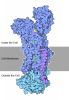


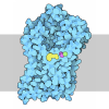





- Assembly
| Deposited unit | 
|
|---|---|
| 1 |
|
-Components
-Guanine nucleotide-binding protein ... , 3 types, 3 molecules ABG
| #1: Protein | Mass: 28362.049 Da / Num. of mol.: 1 / Mutation: G49D, E50N, A249D, S252D, L272D, I372A, V375I Source method: isolated from a genetically manipulated source Source: (gene. exp.) Homo sapiens (human) / Gene: GNAS, GNAS1, GSP / Production host:   Spodoptera frugiperda (fall armyworm) Spodoptera frugiperda (fall armyworm) |
|---|---|
| #2: Protein | Mass: 39728.426 Da / Num. of mol.: 1 Source method: isolated from a genetically manipulated source Details: Guanine nucleotide-binding protein G(I)/G(S)/G(T) subunit beta-1, with C-terminal smBIT tag Source: (gene. exp.) Homo sapiens (human) / Gene: GNB1 / Production host: Spodoptera frugiperda (fall armyworm) / References: UniProt: P62873 |
| #3: Protein | Mass: 7675.814 Da / Num. of mol.: 1 Source method: isolated from a genetically manipulated source Source: (gene. exp.) Homo sapiens (human) / Gene: GNG2 / Production host: Spodoptera frugiperda (fall armyworm) / References: UniProt: P59768 |
-Protein , 1 types, 1 molecules R
| #4: Protein | Mass: 66988.383 Da / Num. of mol.: 1 / Mutation: M7W,H102I,R106L Source method: isolated from a genetically manipulated source Source: (gene. exp.) Escherichia coli (E. coli), (gene. exp.) Homo sapiens (human)Gene: cybC, ADORA2B / Production host: Spodoptera frugiperda (fall armyworm) / References: UniProt: P0ABE7, UniProt: P29275 |
|---|
-Non-polymers , 3 types, 9 molecules 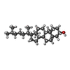
| #5: Chemical | ChemComp-A1EUU / ( Mass: 505.322 Da / Num. of mol.: 1 / Source method: obtained synthetically / Formula: C20H21BrN6O5 / Feature type: SUBJECT OF INVESTIGATION | ||
|---|---|---|---|
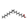
| #6: Chemical | ChemComp-CLR / 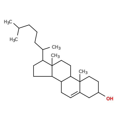 Mass: 386.654 Da / Num. of mol.: 5 / Source method: obtained synthetically / Formula: C27H46O Mass: 386.654 Da / Num. of mol.: 5 / Source method: obtained synthetically / Formula: C27H46O#7: Chemical | 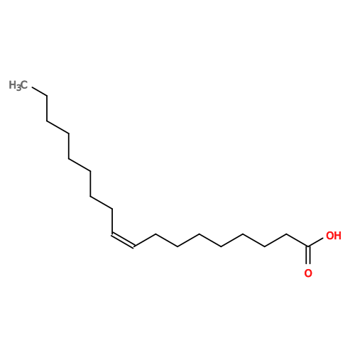 Mass: 282.461 Da / Num. of mol.: 3 / Source method: obtained synthetically / Formula: C18H34O2 Mass: 282.461 Da / Num. of mol.: 3 / Source method: obtained synthetically / Formula: C18H34O2 |
-Details
| Has ligand of interest | Y |
|---|---|
| Has protein modification | Y |
-Experimental details
-Experiment
| Experiment | Method: ELECTRON MICROSCOPY |
|---|---|
| EM experiment | Aggregation state: PARTICLE / 3D reconstruction method: single particle reconstruction |
- Sample preparation
| Component | Name: hA2BR-Gs complex / Type: COMPLEX / Entity ID: #1-#4 / Source: RECOMBINANT |
|---|---|
| Source (natural) | Organism: Homo sapiens (human) |
| Source (recombinant) | Organism: Spodoptera frugiperda (fall armyworm) |
| Buffer solution | pH: 7.5 |
| Specimen | Embedding applied: NO / Shadowing applied: NO / Staining applied: NO / Vitrification applied: YES |
| Vitrification | Cryogen name: ETHANE |
- Electron microscopy imaging
| Experimental equipment |  Model: Titan Krios / Image courtesy: FEI Company |
|---|---|
| Microscopy | Model: TFS KRIOS |
| Electron gun | Electron source:  FIELD EMISSION GUN / Accelerating voltage: 300 kV / Illumination mode: FLOOD BEAM FIELD EMISSION GUN / Accelerating voltage: 300 kV / Illumination mode: FLOOD BEAM |
| Electron lens | Mode: BRIGHT FIELD / Nominal defocus max: 2000 nm / Nominal defocus min: 1000 nm |
| Image recording | Electron dose: 60 e/Å2 / Film or detector model: GATAN K3 (6k x 4k) |
- Processing
| EM software | Name: PHENIX / Version: 1.20.1_4487 / Category: model refinement | ||||||||||||||||||||||||
|---|---|---|---|---|---|---|---|---|---|---|---|---|---|---|---|---|---|---|---|---|---|---|---|---|---|
| CTF correction | Type: PHASE FLIPPING AND AMPLITUDE CORRECTION | ||||||||||||||||||||||||
| 3D reconstruction | Resolution: 3.2 Å / Resolution method: FSC 0.143 CUT-OFF / Num. of particles: 186819 / Symmetry type: POINT | ||||||||||||||||||||||||
| Refinement | Highest resolution: 3.2 Å Stereochemistry target values: REAL-SPACE (WEIGHTED MAP SUM AT ATOM CENTERS) | ||||||||||||||||||||||||
| Refine LS restraints |
|
