 Movie
Movie Controller

[English] 日本語
 Yorodumi
- PDB-9vsm: Crystal structure of cystathionine gamma-synthase from Lactobacil... -
+ Open data

- Basic information
| Entry | Database: PDB / ID: 9vsm | ||||||
|---|---|---|---|---|---|---|---|
| Title | Crystal structure of cystathionine gamma-synthase from Lactobacillus plantarum complexed with the cystathionine-bound external aldimine | ||||||
 Components Components | Cysteine gamma synthase/O-succinylhomoserine (Thiol)-lyase | ||||||
 Keywords Keywords | TRANSFERASE / cystathionine gamma-synthase | ||||||
| Function / homology |  Function and homology information Function and homology informationcystathionine gamma-synthase / carbon-sulfur lyase activity / cystathionine gamma-synthase activity / transsulfuration / pyridoxal phosphate binding / cytoplasm Similarity search - Function | ||||||
| Biological species |  Lactiplantibacillus plantarum (bacteria) Lactiplantibacillus plantarum (bacteria) | ||||||
| Method |  X-RAY DIFFRACTION / SYNCHROTRON / MOLECULAR REPLACEMENT / Resolution: 1.58 Å X-RAY DIFFRACTION / SYNCHROTRON / MOLECULAR REPLACEMENT / Resolution: 1.58 Å | ||||||
 Authors Authors | Oda, K. / Matoba, Y. | ||||||
| Funding support | 1items
| ||||||
 Citation Citation | Journal: Febs Lett. / Year: 2026 Title: Structural insight into the substrate specificity of cystathionine gamma-synthase from Lactobacillus plantarum. Authors: Matoba, Y. / Oda, K. / Ohtani, M. / Mende, Y. / Noda, K. | ||||||
| History |
|
- Structure visualization
| Structure viewer | Molecule: MolmilJmol/JSmol |
|---|
- Downloads & links
-Download
| PDBx/mmCIF format | 9vsm.cif.gz | 327.1 KB | Display | PDBx/mmCIF format |
|---|---|---|---|---|
| PDB format | pdb9vsm.ent.gz | 263.4 KB | Display | PDB format |
| PDBx/mmJSON format | 9vsm.json.gz | Tree view | PDBx/mmJSON format | |
| Others |  Other downloads Other downloads |
-Validation report
| Arichive directory | https://data.pdbj.org/pub/pdb/validation_reports/vs/9vsmftp://data.pdbj.org/pub/pdb/validation_reports/vs/9vsm | HTTPS FTP |
|---|
-Related structure data
| Similar structure data |
|---|
-Links
 PDBj
PDBj- Assembly
| Deposited unit | 
| ||||||||
|---|---|---|---|---|---|---|---|---|---|
| 1 | 
| ||||||||
| 2 | 
| ||||||||
| Unit cell |
| ||||||||
| Components on special symmetry positions |
|
-Components
| #1: Protein | Mass: 41566.707 Da / Num. of mol.: 4 / Mutation: K203A Source method: isolated from a genetically manipulated source Details: This protein was C-terminally fused with histidine tag. Source: (gene. exp.) Lactiplantibacillus plantarum (strain ATCC BAA-793 / NCIMB 8826 / WCFS1) (bacteria)Gene: cgs, lp_2634 / Plasmid: plasmid / Details (production host): pET21 / Production host: #2: Chemical | ChemComp-E9U / ( 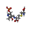  Mass: 451.389 Da / Num. of mol.: 4 / Source method: obtained synthetically / Formula: C15H22N3O9PS / Feature type: SUBJECT OF INVESTIGATION Mass: 451.389 Da / Num. of mol.: 4 / Source method: obtained synthetically / Formula: C15H22N3O9PS / Feature type: SUBJECT OF INVESTIGATION#3: Water | ChemComp-HOH / |  Mass: 18.015 Da / Num. of mol.: 1500 / Source method: isolated from a natural source / Formula: H2O Mass: 18.015 Da / Num. of mol.: 1500 / Source method: isolated from a natural source / Formula: H2OHas ligand of interest | Y | Has protein modification | N | |
|---|
-Experimental details
-Experiment
| Experiment | Method: X-RAY DIFFRACTION / Number of used crystals: 1 |
|---|
- Sample preparation
| Crystal | Density Matthews: 2.58 Å3/Da / Density % sol: 52.41 % |
|---|---|
| Crystal grow | Temperature: 298 K / Method: vapor diffusion, sitting drop / pH: 4 / Details: PEG 4000 |
-Data collection
| Diffraction | Mean temperature: 100 K / Serial crystal experiment: N |
|---|---|
| Diffraction source | Source: SYNCHROTRON / Site: SPring-8  / Beamline: BL44XU / Wavelength: 0.9 Å / Beamline: BL44XU / Wavelength: 0.9 Å |
| Detector | Type: DECTRIS EIGER X 16M / Detector: PIXEL / Date: Apr 21, 2022 |
| Radiation | Protocol: SINGLE WAVELENGTH / Monochromatic (M) / Laue (L): M / Scattering type: x-ray |
| Radiation wavelength | Wavelength: 0.9 Å / Relative weight: 1 |
| Reflection | Resolution: 1.58→48.36 Å / Num. obs: 230342 / % possible obs: 99.9 % / Redundancy: 7.1 % / CC1/2: 0.995 / Rmerge(I) obs: 0.098 / Rpim(I) all: 0.04 / Rrim(I) all: 0.106 / Χ2: 1.14 / Net I/σ(I): 13 / Num. measured all: 1630623 |
| Reflection shell | Resolution: 1.58→1.61 Å / % possible obs: 100 % / Redundancy: 7.2 % / Rmerge(I) obs: 0.455 / Num. measured all: 82272 / Num. unique obs: 11380 / CC1/2: 0.941 / Rpim(I) all: 0.182 / Rrim(I) all: 0.491 / Χ2: 0.91 / Net I/σ(I) obs: 5.1 |
- Processing
| Software |
| |||||||||||||||||||||||||||||||||||||||||||||||||||||||||||||||||||||||||||||||||||||||||||||||||||||||||||||||||||||||||||||||||||||||||||||||||||||||||||||||||||||||||||||||||||||||||||||||||||||||||||||||||||||||||
|---|---|---|---|---|---|---|---|---|---|---|---|---|---|---|---|---|---|---|---|---|---|---|---|---|---|---|---|---|---|---|---|---|---|---|---|---|---|---|---|---|---|---|---|---|---|---|---|---|---|---|---|---|---|---|---|---|---|---|---|---|---|---|---|---|---|---|---|---|---|---|---|---|---|---|---|---|---|---|---|---|---|---|---|---|---|---|---|---|---|---|---|---|---|---|---|---|---|---|---|---|---|---|---|---|---|---|---|---|---|---|---|---|---|---|---|---|---|---|---|---|---|---|---|---|---|---|---|---|---|---|---|---|---|---|---|---|---|---|---|---|---|---|---|---|---|---|---|---|---|---|---|---|---|---|---|---|---|---|---|---|---|---|---|---|---|---|---|---|---|---|---|---|---|---|---|---|---|---|---|---|---|---|---|---|---|---|---|---|---|---|---|---|---|---|---|---|---|---|---|---|---|---|---|---|---|---|---|---|---|---|---|---|---|---|---|---|---|---|
| Refinement | Method to determine structure: MOLECULAR REPLACEMENT / Resolution: 1.58→46.79 Å / SU ML: 0.12 / Cross valid method: FREE R-VALUE / σ(F): 1.36 / Phase error: 16.24 / Stereochemistry target values: ML
| |||||||||||||||||||||||||||||||||||||||||||||||||||||||||||||||||||||||||||||||||||||||||||||||||||||||||||||||||||||||||||||||||||||||||||||||||||||||||||||||||||||||||||||||||||||||||||||||||||||||||||||||||||||||||
| Solvent computation | Shrinkage radii: 0.9 Å / VDW probe radii: 1.11 Å / Solvent model: FLAT BULK SOLVENT MODEL | |||||||||||||||||||||||||||||||||||||||||||||||||||||||||||||||||||||||||||||||||||||||||||||||||||||||||||||||||||||||||||||||||||||||||||||||||||||||||||||||||||||||||||||||||||||||||||||||||||||||||||||||||||||||||
| Refinement step | Cycle: LAST / Resolution: 1.58→46.79 Å
| |||||||||||||||||||||||||||||||||||||||||||||||||||||||||||||||||||||||||||||||||||||||||||||||||||||||||||||||||||||||||||||||||||||||||||||||||||||||||||||||||||||||||||||||||||||||||||||||||||||||||||||||||||||||||
| Refine LS restraints |
| |||||||||||||||||||||||||||||||||||||||||||||||||||||||||||||||||||||||||||||||||||||||||||||||||||||||||||||||||||||||||||||||||||||||||||||||||||||||||||||||||||||||||||||||||||||||||||||||||||||||||||||||||||||||||
| LS refinement shell |
|
