 Movie
Movie Controller

[English] 日本語
 Yorodumi
- PDB-9vpz: Crystal structure of wild-type Trypanosoma brucei DHODH in FMN-re... -
+ Open data

- Basic information
| Entry | Database: PDB / ID: 9vpz | ||||||
|---|---|---|---|---|---|---|---|
| Title | Crystal structure of wild-type Trypanosoma brucei DHODH in FMN-reduced, ligand-free form | ||||||
 Components Components | Dihydroorotate dehydrogenase (fumarate) | ||||||
 Keywords Keywords | FLAVOPROTEIN / Ping-Pong Reaction Mechanism / Substrate/Product-enzyme interactions. | ||||||
| Function / homology |  Function and homology information Function and homology informationdihydroorotate dehydrogenase (fumarate) / dihydroorotate dehydrogenase (fumarate) activity / fumarate metabolic process / dihydroorotate dehydrogenase activity / glycosome / ciliary plasm / 'de novo' UMP biosynthetic process / 'de novo' pyrimidine nucleobase biosynthetic process / nucleoplasm / cytoplasm Similarity search - Function | ||||||
| Biological species |  | ||||||
| Method |  X-RAY DIFFRACTION / SYNCHROTRON / MOLECULAR REPLACEMENT / Resolution: 1.75 Å X-RAY DIFFRACTION / SYNCHROTRON / MOLECULAR REPLACEMENT / Resolution: 1.75 Å | ||||||
 Authors Authors | Kubota, T. / Tani, O. / Yamasaki, K. | ||||||
| Funding support | 1items
| ||||||
 Citation Citation | Journal: J.Mol.Biol. / Year: 2025 Title: Structural Basis of Redox-Dependent Affinities of Dihydroorotate Dehydrogenase for Its Substrates and Products. Authors: Tani, O. / Kubota, T. / Yamasaki, T. / Hirokawa, T. / Furukawa, K. / Yamasaki, K. | ||||||
| History |
|
- Structure visualization
| Structure viewer | Molecule: MolmilJmol/JSmol |
|---|
- Downloads & links
-Download
| PDBx/mmCIF format | 9vpz.cif.gz | 255 KB | Display | PDBx/mmCIF format |
|---|---|---|---|---|
| PDB format | pdb9vpz.ent.gz | 204.9 KB | Display | PDB format |
| PDBx/mmJSON format | 9vpz.json.gz | Tree view | PDBx/mmJSON format | |
| Others |  Other downloads Other downloads |
-Validation report
| Arichive directory | https://data.pdbj.org/pub/pdb/validation_reports/vp/9vpzftp://data.pdbj.org/pub/pdb/validation_reports/vp/9vpz | HTTPS FTP |
|---|
-Related structure data
| Related structure data |  9vprC  9vpsC  9vptC  9vpuC  9vpvC  9vpwC  9vpxC  9vpyC  9vq0C 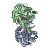 5xfvS S: Starting model for refinement C: citing same article ( |
|---|---|
| Similar structure data |
-Links
 PDBj
PDBj
- Assembly
| Deposited unit | 
| ||||||||
|---|---|---|---|---|---|---|---|---|---|
| 1 | 
| ||||||||
| 2 | 
| ||||||||
| Unit cell |
| ||||||||
| Components on special symmetry positions |
|
-Components
-Protein , 1 types, 4 molecules ACBD
| #1: Protein | Mass: 34473.656 Da / Num. of mol.: 4 / Mutation: A115V Source method: isolated from a genetically manipulated source Source: (gene. exp.) Gene: Tb927.5.3830 Production host:  References: UniProt: Q57U83, dihydroorotate dehydrogenase (fumarate) |
|---|
-Non-polymers , 5 types, 455 molecules 


| #2: Chemical | ChemComp-FNR /  Mass: 458.360 Da / Num. of mol.: 4 / Source method: obtained synthetically / Formula: C17H23N4O9P / Feature type: SUBJECT OF INVESTIGATION Mass: 458.360 Da / Num. of mol.: 4 / Source method: obtained synthetically / Formula: C17H23N4O9P / Feature type: SUBJECT OF INVESTIGATION#3: Chemical | ChemComp-MLI /  Mass: 102.046 Da / Num. of mol.: 11 / Source method: isolated from a natural source / Formula: C3H2O4 Mass: 102.046 Da / Num. of mol.: 11 / Source method: isolated from a natural source / Formula: C3H2O4#4: Chemical | ChemComp-SO2 /  Mass: 64.064 Da / Num. of mol.: 4 / Source method: obtained synthetically / Formula: O2S Mass: 64.064 Da / Num. of mol.: 4 / Source method: obtained synthetically / Formula: O2S#5: Chemical | ChemComp-SO3 /  Mass: 80.063 Da / Num. of mol.: 4 / Source method: obtained synthetically / Formula: SO3 Mass: 80.063 Da / Num. of mol.: 4 / Source method: obtained synthetically / Formula: SO3#6: Water | ChemComp-HOH / | Mass: 18.015 Da / Num. of mol.: 432 / Source method: isolated from a natural source / Formula: H2O |
|---|
-Details
| Has ligand of interest | Y |
|---|---|
| Has protein modification | N |
-Experimental details
-Experiment
| Experiment | Method: X-RAY DIFFRACTION / Number of used crystals: 1 |
|---|
- Sample preparation
| Crystal | Density Matthews: 2.43 Å3/Da / Density % sol: 49.39 % |
|---|---|
| Crystal grow | Temperature: 293 K / Method: vapor diffusion, hanging drop / pH: 6 Details: 1.1 M malonate, 48 mM citrate. soaked into saturated dithionite (< 1min) |
-Data collection
| Diffraction | Mean temperature: 95 K / Serial crystal experiment: N |
|---|---|
| Diffraction source | Source: SYNCHROTRON / Site: Photon Factory  / Beamline: BL-5A / Wavelength: 1 Å / Beamline: BL-5A / Wavelength: 1 Å |
| Detector | Type: DECTRIS PILATUS 2M / Detector: PIXEL / Date: Mar 16, 2018 |
| Radiation | Protocol: SINGLE WAVELENGTH / Monochromatic (M) / Laue (L): M / Scattering type: x-ray |
| Radiation wavelength | Wavelength: 1 Å / Relative weight: 1 |
| Reflection | Resolution: 1.75→50 Å / Num. obs: 136532 / % possible obs: 100 % / Redundancy: 6.58 % / Rmerge(I) obs: 0.086 / Net I/σ(I): 17.6 |
| Reflection shell | Resolution: 1.75→1.78 Å / Rmerge(I) obs: 0.696 / Num. unique obs: 6772 |
- Processing
| Software |
| ||||||||||||||||||||||||||||||||||||||||||||||||||||||||||||||||||||||||||||||||||||||||||||||||||||||||||||||||||||||||||||||||||||||||||||||||||||||||||||||||||||||||||||||||||||||
|---|---|---|---|---|---|---|---|---|---|---|---|---|---|---|---|---|---|---|---|---|---|---|---|---|---|---|---|---|---|---|---|---|---|---|---|---|---|---|---|---|---|---|---|---|---|---|---|---|---|---|---|---|---|---|---|---|---|---|---|---|---|---|---|---|---|---|---|---|---|---|---|---|---|---|---|---|---|---|---|---|---|---|---|---|---|---|---|---|---|---|---|---|---|---|---|---|---|---|---|---|---|---|---|---|---|---|---|---|---|---|---|---|---|---|---|---|---|---|---|---|---|---|---|---|---|---|---|---|---|---|---|---|---|---|---|---|---|---|---|---|---|---|---|---|---|---|---|---|---|---|---|---|---|---|---|---|---|---|---|---|---|---|---|---|---|---|---|---|---|---|---|---|---|---|---|---|---|---|---|---|---|---|---|
| Refinement | Method to determine structure: MOLECULAR REPLACEMENT Starting model: 5XFV Resolution: 1.75→48.88 Å / Cor.coef. Fo:Fc: 0.96 / Cor.coef. Fo:Fc free: 0.946 / SU B: 2.508 / SU ML: 0.078 / Cross valid method: THROUGHOUT / ESU R: 0.109 / ESU R Free: 0.101 / Stereochemistry target values: MAXIMUM LIKELIHOOD / Details: HYDROGENS HAVE BEEN ADDED IN THE RIDING POSITIONS
| ||||||||||||||||||||||||||||||||||||||||||||||||||||||||||||||||||||||||||||||||||||||||||||||||||||||||||||||||||||||||||||||||||||||||||||||||||||||||||||||||||||||||||||||||||||||
| Solvent computation | Ion probe radii: 0.8 Å / Shrinkage radii: 0.8 Å / VDW probe radii: 1.2 Å / Solvent model: MASK | ||||||||||||||||||||||||||||||||||||||||||||||||||||||||||||||||||||||||||||||||||||||||||||||||||||||||||||||||||||||||||||||||||||||||||||||||||||||||||||||||||||||||||||||||||||||
| Displacement parameters | Biso mean: 20.888 Å2
| ||||||||||||||||||||||||||||||||||||||||||||||||||||||||||||||||||||||||||||||||||||||||||||||||||||||||||||||||||||||||||||||||||||||||||||||||||||||||||||||||||||||||||||||||||||||
| Refinement step | Cycle: 1 / Resolution: 1.75→48.88 Å
| ||||||||||||||||||||||||||||||||||||||||||||||||||||||||||||||||||||||||||||||||||||||||||||||||||||||||||||||||||||||||||||||||||||||||||||||||||||||||||||||||||||||||||||||||||||||
| Refine LS restraints |
|