 Movie
Movie Controller

[English] 日本語
 Yorodumi
- PDB-9vfz: Crystal structure of Phaeodactylibacter sp. phosphoglucomutase in... -
+ Open data

- Basic information
| Entry | Database: PDB / ID: 9vfz | |||||||||
|---|---|---|---|---|---|---|---|---|---|---|
| Title | Crystal structure of Phaeodactylibacter sp. phosphoglucomutase in complex with glucose-1-phosphate | |||||||||
 Components Components | Phosphoglucomutase (Alpha-D-glucose-1,6-bisphosphate-dependent) | |||||||||
 Keywords Keywords | ISOMERASE / Metal-binding / Phosphoserine | |||||||||
| Function / homology | 1-O-phosphono-alpha-D-glucopyranose / :  Function and homology information Function and homology information | |||||||||
| Biological species |  Phaeodactylibacter sp. (bacteria) Phaeodactylibacter sp. (bacteria) | |||||||||
| Method |  X-RAY DIFFRACTION / SYNCHROTRON / MOLECULAR REPLACEMENT / Resolution: 1.95 Å X-RAY DIFFRACTION / SYNCHROTRON / MOLECULAR REPLACEMENT / Resolution: 1.95 Å | |||||||||
 Authors Authors | Shen, Y.W. / Tu, T. | |||||||||
| Funding support |  China, 2items China, 2items
| |||||||||
 Citation Citation | Journal: To Be Published Title: In Vitro Design and Optimization of a High-Efficiency Artificial Enzymatic Pathway for Cellulose-to-Starch Conversion Authors: Shen, Y.W. / Tu, T. | |||||||||
| History |
|
- Structure visualization
| Structure viewer | Molecule: MolmilJmol/JSmol |
|---|
- Downloads & links
-Download
| PDBx/mmCIF format | 9vfz.cif.gz | 380.6 KB | Display | PDBx/mmCIF format |
|---|---|---|---|---|
| PDB format | pdb9vfz.ent.gz | 260.9 KB | Display | PDB format |
| PDBx/mmJSON format | 9vfz.json.gz | Tree view | PDBx/mmJSON format | |
| Others |  Other downloads Other downloads |
-Validation report
| Arichive directory | https://data.pdbj.org/pub/pdb/validation_reports/vf/9vfzftp://data.pdbj.org/pub/pdb/validation_reports/vf/9vfz | HTTPS FTP |
|---|
-Related structure data


-Links
 PDBj
PDBj- Assembly
| Deposited unit | 
| ||||||||||||
|---|---|---|---|---|---|---|---|---|---|---|---|---|---|
| 1 |
| ||||||||||||
| Unit cell |
|
-Components
| #1: Protein | Mass: 64126.938 Da / Num. of mol.: 1 Source method: isolated from a genetically manipulated source Source: (gene. exp.) Phaeodactylibacter sp. (bacteria) / Gene: pgm, KDD09_20305 / Production host: References: UniProt: A0A959BHL9, phosphoglucomutase (alpha-D-glucose-1,6-bisphosphate-dependent) |
|---|---|
| #2: Chemical | ChemComp-MG /   Mass: 24.305 Da / Num. of mol.: 1 / Source method: obtained synthetically / Formula: Mg / Feature type: SUBJECT OF INVESTIGATION Mass: 24.305 Da / Num. of mol.: 1 / Source method: obtained synthetically / Formula: Mg / Feature type: SUBJECT OF INVESTIGATION |
| #3: Sugar | ChemComp-G1P / 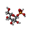  Type: D-saccharide / Mass: 260.136 Da / Num. of mol.: 1 / Source method: obtained synthetically / Formula: C6H13O9P / Feature type: SUBJECT OF INVESTIGATION Type: D-saccharide / Mass: 260.136 Da / Num. of mol.: 1 / Source method: obtained synthetically / Formula: C6H13O9P / Feature type: SUBJECT OF INVESTIGATION |
| #4: Water | ChemComp-HOH /  Mass: 18.015 Da / Num. of mol.: 53 / Source method: isolated from a natural source / Formula: H2O Mass: 18.015 Da / Num. of mol.: 53 / Source method: isolated from a natural source / Formula: H2O |
| Has ligand of interest | Y |
| Has protein modification | N |
-Experimental details
-Experiment
| Experiment | Method: X-RAY DIFFRACTION / Number of used crystals: 1 |
|---|
- Sample preparation
| Crystal | Density Matthews: 2.2 Å3/Da / Density % sol: 44.01 % |
|---|---|
| Crystal grow | Temperature: 289.15 K / Method: vapor diffusion, hanging drop Details: 0.15 M DL-maleic acid pH 7.0 0.1 M Imidazole pH 7.0 24% v/v polyethylene glycol monomethyl ether 550 3 mM G1P |
-Data collection
| Diffraction | Mean temperature: 100 K / Serial crystal experiment: N |
|---|---|
| Diffraction source | Source: SYNCHROTRON / Site: SSRF / Beamline: BL19U1 / Wavelength: 0.97861 Å |
| Detector | Type: DECTRIS PILATUS3 6M / Detector: PIXEL / Date: Apr 4, 2025 |
| Radiation | Protocol: SINGLE WAVELENGTH / Monochromatic (M) / Laue (L): M / Scattering type: x-ray |
| Radiation wavelength | Wavelength: 0.97861 Å / Relative weight: 1 |
| Reflection | Resolution: 1.95→50 Å / Num. obs: 40136 / % possible obs: 99.3 % / Redundancy: 6.48 % / CC1/2: 0.9349 / Net I/σ(I): 8.14 |
| Reflection shell | Resolution: 1.95→2 Å / Num. unique obs: 2967 / CC1/2: 0.6943 / % possible all: 99.7 |
- Processing
| Software |
| |||||||||||||||||||||||||||||||||||||||||||||||||||||||||||||||||||||||||||||||||||||||||||||||||||||||||
|---|---|---|---|---|---|---|---|---|---|---|---|---|---|---|---|---|---|---|---|---|---|---|---|---|---|---|---|---|---|---|---|---|---|---|---|---|---|---|---|---|---|---|---|---|---|---|---|---|---|---|---|---|---|---|---|---|---|---|---|---|---|---|---|---|---|---|---|---|---|---|---|---|---|---|---|---|---|---|---|---|---|---|---|---|---|---|---|---|---|---|---|---|---|---|---|---|---|---|---|---|---|---|---|---|---|---|
| Refinement | Method to determine structure: MOLECULAR REPLACEMENT / Resolution: 1.95→49.16 Å / SU ML: 0.3742 / Cross valid method: NONE / σ(F): 1.35 / Phase error: 46.5958 / Stereochemistry target values: CDL v1.2
| |||||||||||||||||||||||||||||||||||||||||||||||||||||||||||||||||||||||||||||||||||||||||||||||||||||||||
| Solvent computation | Shrinkage radii: 0.9 Å / VDW probe radii: 1.11 Å / Solvent model: FLAT BULK SOLVENT MODEL | |||||||||||||||||||||||||||||||||||||||||||||||||||||||||||||||||||||||||||||||||||||||||||||||||||||||||
| Displacement parameters | Biso mean: 64.03 Å2 | |||||||||||||||||||||||||||||||||||||||||||||||||||||||||||||||||||||||||||||||||||||||||||||||||||||||||
| Refinement step | Cycle: LAST / Resolution: 1.95→49.16 Å
| |||||||||||||||||||||||||||||||||||||||||||||||||||||||||||||||||||||||||||||||||||||||||||||||||||||||||
| Refine LS restraints |
| |||||||||||||||||||||||||||||||||||||||||||||||||||||||||||||||||||||||||||||||||||||||||||||||||||||||||
| LS refinement shell |
| |||||||||||||||||||||||||||||||||||||||||||||||||||||||||||||||||||||||||||||||||||||||||||||||||||||||||
| Refinement TLS params. | Method: refined / Origin x: 10.0141877041 Å / Origin y: -18.096012326 Å / Origin z: 23.006211665 Å
| |||||||||||||||||||||||||||||||||||||||||||||||||||||||||||||||||||||||||||||||||||||||||||||||||||||||||
| Refinement TLS group | Selection details: all |
