 Movie
Movie Controller


+ Open data

- Basic information
| Entry | Database: PDB / ID: 9vfq | ||||||||||||||||||||||||
|---|---|---|---|---|---|---|---|---|---|---|---|---|---|---|---|---|---|---|---|---|---|---|---|---|---|
| Title | Structure of mature CVA6 virus | ||||||||||||||||||||||||
 Components Components |
| ||||||||||||||||||||||||
 Keywords Keywords | VIRUS / coxsackievirus A6 / KRM1 / receptor | ||||||||||||||||||||||||
| Function / homology |  Function and homology information Function and homology informationsymbiont-mediated suppression of host cytoplasmic pattern recognition receptor signaling pathway via inhibition of MDA-5 activity / picornain 2A / symbiont-mediated suppression of host mRNA export from nucleus / symbiont genome entry into host cell via pore formation in plasma membrane / picornain 3C / T=pseudo3 icosahedral viral capsid / host cell cytoplasmic vesicle membrane / virion component / viral capsid / ribonucleoside triphosphate phosphatase activity ...symbiont-mediated suppression of host cytoplasmic pattern recognition receptor signaling pathway via inhibition of MDA-5 activity / picornain 2A / symbiont-mediated suppression of host mRNA export from nucleus / symbiont genome entry into host cell via pore formation in plasma membrane / picornain 3C / T=pseudo3 icosahedral viral capsid / host cell cytoplasmic vesicle membrane / virion component / viral capsid / ribonucleoside triphosphate phosphatase activity / host cell / nucleoside-triphosphate phosphatase / channel activity / monoatomic ion transmembrane transport / DNA replication / RNA helicase activity / endocytosis involved in viral entry into host cell / symbiont-mediated suppression of host gene expression / symbiont-mediated activation of host autophagy / RNA-directed RNA polymerase / cysteine-type endopeptidase activity / viral RNA genome replication / RNA-directed RNA polymerase activity / symbiont entry into host cell / DNA-templated transcription / virion attachment to host cell / host cell nucleus / structural molecule activity / proteolysis / RNA binding / zinc ion binding / ATP binding Similarity search - Function | ||||||||||||||||||||||||
| Biological species |  Coxsackievirus A6 Coxsackievirus A6 | ||||||||||||||||||||||||
| Method | ELECTRON MICROSCOPY / single particle reconstruction / cryo EM / Resolution: 2.23 Å | ||||||||||||||||||||||||
 Authors Authors | Ke, X. / Li, X. / Liu, Z. / Liu, K. / Yan, X. / Shu, B. / Zhang, C. | ||||||||||||||||||||||||
| Funding support |  China, 1items China, 1items
| ||||||||||||||||||||||||
 Citation Citation | Journal: Nat Commun / Year: 2025 Title: Molecular mechanisms of receptor recognition and antibody neutralization of coxsackievirus A6. Authors: Xianliang Ke / Xue Li / Zeyu Liu / Kexin Liu / Weichi Liu / Xingyu Yan / Bo Shu / Chao Zhang / Abstract: Coxsackievirus A6 (CVA6), a major cause of hand, foot, and mouth disease, lacks approved vaccines or drugs. KRM1 is its only known receptor, but its precise role remains unclear. This study ...Coxsackievirus A6 (CVA6), a major cause of hand, foot, and mouth disease, lacks approved vaccines or drugs. KRM1 is its only known receptor, but its precise role remains unclear. This study investigates CVA6's entry mechanism and antibody neutralization. Cryo-EM shows CVA6 clinical strain HeB primarily exists as mature virions. KRM1 binding within the canyon triggers conversion to uncoating intermediate, defining KRM1 as an uncoating receptor for CVA6. However, KRM1 knockout reduces CVA6 infectivity without affecting attachment. Conversely, disrupting heparan sulfate proteoglycan (HSPG) impairs both viral attachment and infectivity, and CVA6 virions bind heparin directly. These results support a two-receptor entry model for CVA6: HSPG mediates viral attachment, while KRM1 induces uncoating. Additionally, we develop two CVA6-specific protective antibodies (1F4 and 3H7), targeting a new antigenic site near the three-fold axis of the viral capsid. These antibodies sterically block KRM1 binding and function post-attachment, consistent with KRM1's role. The findings elucidate CVA6 entry and offer a basis for antibody interventions. | ||||||||||||||||||||||||
| History |
|
- Structure visualization
| Structure viewer | Molecule: MolmilJmol/JSmol |
|---|
- Downloads & links
-Download
| PDBx/mmCIF format | 9vfq.cif.gz | 153.2 KB | Display | PDBx/mmCIF format |
|---|---|---|---|---|
| PDB format | pdb9vfq.ent.gz | 118 KB | Display | PDB format |
| PDBx/mmJSON format | 9vfq.json.gz | Tree view | PDBx/mmJSON format | |
| Others |  Other downloads Other downloads |
-Validation report
| Arichive directory | https://data.pdbj.org/pub/pdb/validation_reports/vf/9vfqftp://data.pdbj.org/pub/pdb/validation_reports/vf/9vfq | HTTPS FTP |
|---|
-Related structure data
| Related structure data |  65031MC  9vfpC  9vfrC  9vfsC  9vftC  9vfuC  9vg1C M: map data used to model this data C: citing same article ( |
|---|---|
| Similar structure data |


-Links
 PDBj
PDBj


- Assembly
| Deposited unit | 
|
|---|---|
| 1 | x 60
|
-Components
| #1: Protein | Mass: 33568.379 Da / Num. of mol.: 1 / Source method: isolated from a natural source / Source: (natural) Coxsackievirus A6 / References: UniProt: A0A222NWY2 |
|---|---|
| #2: Protein | Mass: 28138.604 Da / Num. of mol.: 1 / Source method: isolated from a natural source / Source: (natural) Coxsackievirus A6References: UniProt: A0A7D0TR32, picornain 2A, nucleoside-triphosphate phosphatase, picornain 3C, RNA-directed RNA polymerase |
| #3: Protein | Mass: 26324.760 Da / Num. of mol.: 1 / Source method: isolated from a natural source / Source: (natural) Coxsackievirus A6References: UniProt: A0A4P2SK07, picornain 2A, nucleoside-triphosphate phosphatase, picornain 3C, RNA-directed RNA polymerase |
| #4: Protein | Mass: 7503.163 Da / Num. of mol.: 1 / Source method: isolated from a natural source / Source: (natural) Coxsackievirus A6 / References: UniProt: E3VJS6 |
| #5: Chemical | ChemComp-STE / 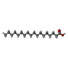  Mass: 284.477 Da / Num. of mol.: 1 / Source method: obtained synthetically / Formula: C18H36O2 / Feature type: SUBJECT OF INVESTIGATION Mass: 284.477 Da / Num. of mol.: 1 / Source method: obtained synthetically / Formula: C18H36O2 / Feature type: SUBJECT OF INVESTIGATION |
| Has ligand of interest | Y |
| Has protein modification | N |
-Experimental details
-Experiment
| Experiment | Method: ELECTRON MICROSCOPY |
|---|---|
| EM experiment | Aggregation state: PARTICLE / 3D reconstruction method: single particle reconstruction |
- Sample preparation
| Component | Name: mature CVA6 virus / Type: COMPLEX / Details: UV inactivated Coxsackievirus A6 (CV-A6) / Entity ID: #1-#4 / Source: NATURAL |
|---|---|
| Source (natural) | Organism: Coxsackievirus A6 |
| Source (recombinant) | Organism:  Homo sapiens (human) Homo sapiens (human) |
| Buffer solution | pH: 7.4 |
| Specimen | Embedding applied: NO / Shadowing applied: NO / Staining applied: NO / Vitrification applied: YES |
| Specimen support | Grid material: COPPER / Grid mesh size: 200 divisions/in. / Grid type: EMS Lacey Carbon |
| Vitrification | Instrument: FEI VITROBOT MARK IV / Cryogen name: ETHANE / Humidity: 100 % / Chamber temperature: 273 K |
- Electron microscopy imaging
| Experimental equipment |  Model: Titan Krios / Image courtesy: FEI Company |
|---|---|
| Microscopy | Model: TFS KRIOS |
| Electron gun | Electron source:  FIELD EMISSION GUN / Accelerating voltage: 300 kV / Illumination mode: FLOOD BEAM FIELD EMISSION GUN / Accelerating voltage: 300 kV / Illumination mode: FLOOD BEAM |
| Electron lens | Mode: BRIGHT FIELD / Nominal defocus max: 2500 nm / Nominal defocus min: 500 nm / Cs: 2.7 mm |
| Specimen holder | Cryogen: NITROGEN / Specimen holder model: FEI TITAN KRIOS AUTOGRID HOLDER |
| Image recording | Electron dose: 50 e/Å2 / Film or detector model: FEI FALCON IV (4k x 4k) |
- Processing
| EM software |
| ||||||||||||||||||||||||
|---|---|---|---|---|---|---|---|---|---|---|---|---|---|---|---|---|---|---|---|---|---|---|---|---|---|
| CTF correction | Type: PHASE FLIPPING AND AMPLITUDE CORRECTION | ||||||||||||||||||||||||
| Symmetry | Point symmetry: I (icosahedral) | ||||||||||||||||||||||||
| 3D reconstruction | Resolution: 2.23 Å / Resolution method: FSC 0.143 CUT-OFF / Num. of particles: 19060 / Algorithm: FOURIER SPACE / Symmetry type: POINT | ||||||||||||||||||||||||
| Refinement | Stereochemistry target values: REAL-SPACE (WEIGHTED MAP SUM AT ATOM CENTERS) | ||||||||||||||||||||||||
| Refine LS restraints |
|
