 Movie
Movie Controller

[English] 日本語
 Yorodumi
- PDB-9uwz: Crystal structure of the type III secretion chaperone VecA from V... -
+ Open data

- Basic information
| Entry | Database: PDB / ID: 9uwz | ||||||
|---|---|---|---|---|---|---|---|
| Title | Crystal structure of the type III secretion chaperone VecA from Vibrio parahaemolyticus | ||||||
 Components Components | CesT family type III secretion system chaperone | ||||||
 Keywords Keywords | CHAPERONE / Vibrio parahaemolyticus / Type 3 secretion system | ||||||
| Function / homology | Tir chaperone protein (CesT) family / Tir chaperone protein (CesT) family / protein secretion by the type III secretion system / PHOSPHATE ION / HEXATANTALUM DODECABROMIDE / CesT family type III secretion system chaperone Function and homology information Function and homology information | ||||||
| Biological species |   Vibrio parahaemolyticus (bacteria) Vibrio parahaemolyticus (bacteria) | ||||||
| Method |  X-RAY DIFFRACTION / SYNCHROTRON / SAD / Resolution: 2.2 Å X-RAY DIFFRACTION / SYNCHROTRON / SAD / Resolution: 2.2 Å | ||||||
 Authors Authors | Iimori, M. / Oki, H. / Akeda, Y. / Ishii, E. / Kodama, T. / Ueda, T. / Nakamura, S. / Matsuda, S. / Kawahara, K. / Iida, T. | ||||||
| Funding support |  Japan, 1items Japan, 1items
| ||||||
 Citation Citation | Journal: Biochem.Biophys.Res.Commun. / Year: 2025 Title: Structural basis of effector recognition by the T3SS chaperone VecA from Vibrio parahaemolyticus. Authors: Iimori, M. / Oki, H. / Akeda, Y. / Ishii, E. / Kodama, T. / Ueda, T. / Nakamura, S. / Matsuda, S. / Kawahara, K. / Iida, T. | ||||||
| History |
|
- Structure visualization
| Structure viewer | Molecule: MolmilJmol/JSmol |
|---|
- Downloads & links
-Download
| PDBx/mmCIF format | 9uwz.cif.gz | 72 KB | Display | PDBx/mmCIF format |
|---|---|---|---|---|
| PDB format | pdb9uwz.ent.gz | 48.3 KB | Display | PDB format |
| PDBx/mmJSON format | 9uwz.json.gz | Tree view | PDBx/mmJSON format | |
| Others |  Other downloads Other downloads |
-Validation report
| Arichive directory | https://data.pdbj.org/pub/pdb/validation_reports/uw/9uwzftp://data.pdbj.org/pub/pdb/validation_reports/uw/9uwz | HTTPS FTP |
|---|
-Related structure data

-Links
 PDBj
PDBj

- Assembly
| Deposited unit | 
| ||||||||||||
|---|---|---|---|---|---|---|---|---|---|---|---|---|---|
| 1 |
| ||||||||||||
| Unit cell |
|
-Components
| #1: Protein | Mass: 17553.850 Da / Num. of mol.: 2 Source method: isolated from a genetically manipulated source Source: (gene. exp.) Vibrio parahaemolyticus (bacteria) / Gene: VP1682 / Production host: #2: Chemical | ChemComp-PO4 / |   Mass: 94.971 Da / Num. of mol.: 1 / Source method: obtained synthetically / Formula: PO4 Mass: 94.971 Da / Num. of mol.: 1 / Source method: obtained synthetically / Formula: PO4#3: Chemical | ChemComp-TBR / | 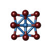  Mass: 2044.535 Da / Num. of mol.: 1 / Source method: isolated from a natural source / Formula: Br12Ta6 Mass: 2044.535 Da / Num. of mol.: 1 / Source method: isolated from a natural source / Formula: Br12Ta6#4: Water | ChemComp-HOH / |  Mass: 18.015 Da / Num. of mol.: 18 / Source method: isolated from a natural source / Formula: H2O Mass: 18.015 Da / Num. of mol.: 18 / Source method: isolated from a natural source / Formula: H2OHas ligand of interest | N | Has protein modification | N | |
|---|
-Experimental details
-Experiment
| Experiment | Method: X-RAY DIFFRACTION / Number of used crystals: 1 |
|---|
- Sample preparation
| Crystal | Density Matthews: 2.13 Å3/Da / Density % sol: 42.22 % |
|---|---|
| Crystal grow | Temperature: 277 K / Method: vapor diffusion, sitting drop / Details: 0.1M Tris-HCl (pH 8.8), 1.8M ammonium sulfate |
-Data collection
| Diffraction | Mean temperature: 100 K / Serial crystal experiment: N |
|---|---|
| Diffraction source | Source: SYNCHROTRON / Site: SPring-8 / Beamline: BL38B1 / Wavelength: 1.2538 Å |
| Detector | Type: DECTRIS PILATUS3 6M / Detector: PIXEL / Date: Nov 7, 2018 |
| Radiation | Protocol: SINGLE WAVELENGTH / Monochromatic (M) / Laue (L): M / Scattering type: x-ray |
| Radiation wavelength | Wavelength: 1.2538 Å / Relative weight: 1 |
| Reflection | Resolution: 2.2→47.12 Å / Num. obs: 16059 / % possible obs: 100 % / Redundancy: 23.8 % / Biso Wilson estimate: 55.74 Å2 / CC1/2: 0.999 / Rmerge(I) obs: 0.12 / Rpim(I) all: 0.025 / Rrim(I) all: 0.122 / Χ2: 1.05 / Net I/σ(I): 20.9 |
| Reflection shell | Resolution: 2.2→2.27 Å / Redundancy: 24.6 % / Rmerge(I) obs: 1.612 / Num. unique obs: 1354 / CC1/2: 0.794 / Rpim(I) all: 0.329 / Rrim(I) all: 1.646 / Χ2: 0.65 |
- Processing
| Software |
| |||||||||||||||||||||||||||||||||||||||||||||||||||||||||||||||||||||||||||||||||||||||||||
|---|---|---|---|---|---|---|---|---|---|---|---|---|---|---|---|---|---|---|---|---|---|---|---|---|---|---|---|---|---|---|---|---|---|---|---|---|---|---|---|---|---|---|---|---|---|---|---|---|---|---|---|---|---|---|---|---|---|---|---|---|---|---|---|---|---|---|---|---|---|---|---|---|---|---|---|---|---|---|---|---|---|---|---|---|---|---|---|---|---|---|---|---|
| Refinement | Method to determine structure: SAD / Resolution: 2.2→32.81 Å / SU ML: 0.2973 / Cross valid method: FREE R-VALUE / σ(F): 1.52 / Phase error: 29.4287 Stereochemistry target values: GeoStd + Monomer Library + CDL v1.2
| |||||||||||||||||||||||||||||||||||||||||||||||||||||||||||||||||||||||||||||||||||||||||||
| Solvent computation | Shrinkage radii: 0.9 Å / VDW probe radii: 1.11 Å / Solvent model: FLAT BULK SOLVENT MODEL | |||||||||||||||||||||||||||||||||||||||||||||||||||||||||||||||||||||||||||||||||||||||||||
| Displacement parameters | Biso mean: 67.97 Å2 | |||||||||||||||||||||||||||||||||||||||||||||||||||||||||||||||||||||||||||||||||||||||||||
| Refinement step | Cycle: LAST / Resolution: 2.2→32.81 Å
| |||||||||||||||||||||||||||||||||||||||||||||||||||||||||||||||||||||||||||||||||||||||||||
| Refine LS restraints |
| |||||||||||||||||||||||||||||||||||||||||||||||||||||||||||||||||||||||||||||||||||||||||||
| LS refinement shell |
|
