 Movie
Movie Controller

[English] 日本語
 Yorodumi
- PDB-9uek: Cryo-EM structure of L-lysine 6-dehydrogenase complex with NAD+ a... -
+ Open data

- Basic information
| Entry | Database: PDB / ID: 9uek | |||||||||
|---|---|---|---|---|---|---|---|---|---|---|
| Title | Cryo-EM structure of L-lysine 6-dehydrogenase complex with NAD+ and L-lysine | |||||||||
 Components Components | Lysine 6-dehydrogenase | |||||||||
 Keywords Keywords | OXIDOREDUCTASE / dehydrogenase / complex / amino acid / enzyme | |||||||||
| Function / homology |  Function and homology information Function and homology informationlysine 6-dehydrogenase / L-lysine 6-dehydrogenase activity / protein homooligomerization Similarity search - Function | |||||||||
| Biological species |   Geobacillus stearothermophilus (bacteria) Geobacillus stearothermophilus (bacteria) | |||||||||
| Method | ELECTRON MICROSCOPY / single particle reconstruction / cryo EM / Resolution: 2.53 Å | |||||||||
 Authors Authors | Funahashi, T. / Yamaguchi, H. / Suzuki, S. / Suzuki, H. / Nishikawa, K. / Kazutoshi, T. / Moemi, T. / Toshimi, M. / Hiroshi, M. / Fujiyoshi, Y. / Sugiki, M. | |||||||||
| Funding support | 1items
| |||||||||
 Citation Citation | Journal: J Struct Biol / Year: 2026 Title: Structural basis for substrate recognition in l-lysine 6-dehydrogenase from Geobacillus stearothermophilus by Cryo-EM. Authors: Toshiya Funahashi / Hiroki Yamaguchi / Shota Suzuki / Hiroshi Suzuki / Kouki Nishikawa / Kazutoshi Takahashi / Moemi Tatsumi / Toshimi Mizukoshi / Hiroshi Miyano / Yoshinori Fujiyoshi / Masayuki Sugiki /  Abstract: l-lysine 6-dehydrogenase (LysDH; EC 1.4.1.18) oxidatively deaminates the ε-amino group of l-lysine. Due to its high substrate specificity, LysDH serves as a valuable tool for l-lysine quantification. ...l-lysine 6-dehydrogenase (LysDH; EC 1.4.1.18) oxidatively deaminates the ε-amino group of l-lysine. Due to its high substrate specificity, LysDH serves as a valuable tool for l-lysine quantification. However, the molecular basis of this specificity has remained unclear because of the lack of substrate-bound structures. In this study, we determined the cryo-electron microscopy (cryo-EM) structures of LysDH from the thermophilic bacterium Geobacillus stearothermophilus (GstLysDH) in the apo form at 2.9 Å resolution and in complex with NAD and l-lysine at 2.5 Å resolution. GstLysDH assembles as a tetramer, which undergoes a global conformational transition upon NAD binding. Structural analysis revealed that the α-carboxyl and α-amino groups of l-lysine were coordinated by oppositely charged residues, thereby orienting the ε-amino group toward the nicotinamide ring of NAD and anchoring the substrate in the optimal binding mode. This precise recognition mechanism accounts for the enzyme's strict specificity for the ε-amino group of l-lysine. Furthermore, comparative structural analysis with l-phenylalanine dehydrogenase suggests that the oxidative deamination in GstLysDH proceeds through a conserved hydride transfer mechanism. Together, these insights establish a structural framework for the rational design and industrial application of LysDH and related amino acid dehydrogenases. | |||||||||
| History |
|
- Structure visualization
| Structure viewer | Molecule: MolmilJmol/JSmol |
|---|
- Downloads & links
-Download
| PDBx/mmCIF format | 9uek.cif.gz | 301.1 KB | Display | PDBx/mmCIF format |
|---|---|---|---|---|
| PDB format | pdb9uek.ent.gz | 244.4 KB | Display | PDB format |
| PDBx/mmJSON format | 9uek.json.gz | Tree view | PDBx/mmJSON format | |
| Others |  Other downloads Other downloads |
-Validation report
| Arichive directory | https://data.pdbj.org/pub/pdb/validation_reports/ue/9uekftp://data.pdbj.org/pub/pdb/validation_reports/ue/9uek | HTTPS FTP |
|---|
-Related structure data
| Related structure data |  64084MC  9uejC M: map data used to model this data C: citing same article ( |
|---|---|
| Similar structure data |

-Links
 PDBj
PDBj- Assembly
| Deposited unit | 
| ||||||||||||||||||||||||||||||||||||||||||||||||||||||||||||||||||||||||||||||||||||||||||||||||||
|---|---|---|---|---|---|---|---|---|---|---|---|---|---|---|---|---|---|---|---|---|---|---|---|---|---|---|---|---|---|---|---|---|---|---|---|---|---|---|---|---|---|---|---|---|---|---|---|---|---|---|---|---|---|---|---|---|---|---|---|---|---|---|---|---|---|---|---|---|---|---|---|---|---|---|---|---|---|---|---|---|---|---|---|---|---|---|---|---|---|---|---|---|---|---|---|---|---|---|---|
| 1 |
| ||||||||||||||||||||||||||||||||||||||||||||||||||||||||||||||||||||||||||||||||||||||||||||||||||
| Noncrystallographic symmetry (NCS) | NCS domain:
NCS domain segments: Component-ID: _ / Beg auth comp-ID: MET / Beg label comp-ID: MET / End auth comp-ID: HIS / End label comp-ID: HIS / Refine code: _ / Auth seq-ID: 1 - 384 / Label seq-ID: 1 - 384
NCS ensembles :
|
-Components
| #1: Protein | Mass: 42210.449 Da / Num. of mol.: 4 Source method: isolated from a genetically manipulated source Source: (gene. exp.) Geobacillus stearothermophilus (bacteria)Gene: lysDH / Production host:  Escherichia coli (E. coli) / References: UniProt: Q9AJC6, lysine 6-dehydrogenase Escherichia coli (E. coli) / References: UniProt: Q9AJC6, lysine 6-dehydrogenase#2: Chemical | ChemComp-NAD / 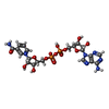 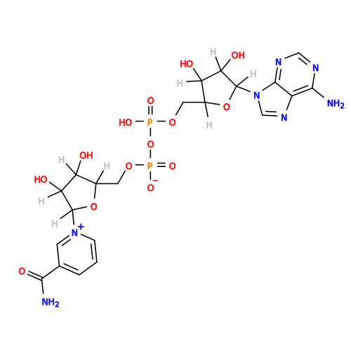 Mass: 663.425 Da / Num. of mol.: 4 / Source method: obtained synthetically / Formula: C21H27N7O14P2 / Feature type: SUBJECT OF INVESTIGATION / Comment: NAD*YM Mass: 663.425 Da / Num. of mol.: 4 / Source method: obtained synthetically / Formula: C21H27N7O14P2 / Feature type: SUBJECT OF INVESTIGATION / Comment: NAD*YM#3: Chemical | ChemComp-LYS / 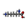 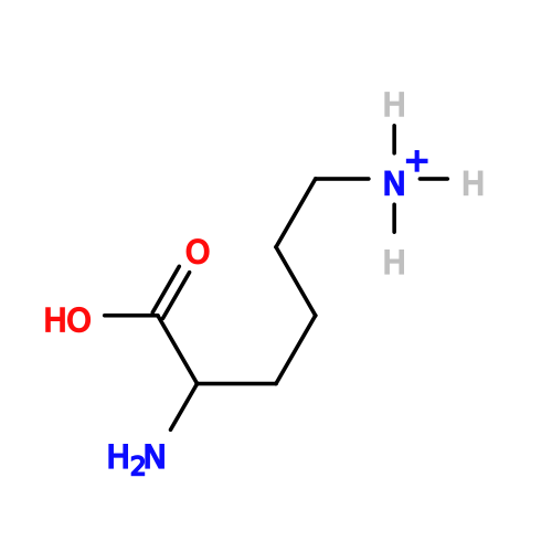 Type: L-peptide linking / Mass: 147.195 Da / Num. of mol.: 4 / Source method: obtained synthetically / Formula: C6H15N2O2 / Feature type: SUBJECT OF INVESTIGATION Type: L-peptide linking / Mass: 147.195 Da / Num. of mol.: 4 / Source method: obtained synthetically / Formula: C6H15N2O2 / Feature type: SUBJECT OF INVESTIGATION#4: Water | ChemComp-HOH / |  Mass: 18.015 Da / Num. of mol.: 32 / Source method: isolated from a natural source / Formula: H2O Mass: 18.015 Da / Num. of mol.: 32 / Source method: isolated from a natural source / Formula: H2OHas ligand of interest | Y | Has protein modification | N | |
|---|
-Experimental details
-Experiment
| Experiment | Method: ELECTRON MICROSCOPY |
|---|---|
| EM experiment | Aggregation state: PARTICLE / 3D reconstruction method: single particle reconstruction |
- Sample preparation
| Component | Name: L-lysine 6-dehydrogenase / Type: COMPLEX / Entity ID: #1 / Source: RECOMBINANT |
|---|---|
| Source (natural) | Organism: Geobacillus stearothermophilus (bacteria) |
| Source (recombinant) | Organism: Escherichia coli (E. coli) |
| Buffer solution | pH: 10 |
| Specimen | Embedding applied: NO / Shadowing applied: NO / Staining applied: NO / Vitrification applied: YES |
| Vitrification | Cryogen name: ETHANE |
- Electron microscopy imaging
| Microscopy | Model: JEOL CRYO ARM 300 |
|---|---|
| Electron gun | Electron source:  FIELD EMISSION GUN / Accelerating voltage: 300 kV / Illumination mode: FLOOD BEAM FIELD EMISSION GUN / Accelerating voltage: 300 kV / Illumination mode: FLOOD BEAM |
| Electron lens | Mode: BRIGHT FIELD / Nominal defocus max: 1500 nm / Nominal defocus min: 800 nm |
| Image recording | Electron dose: 50 e/Å2 / Film or detector model: GATAN K3 (6k x 4k) |
- Processing
| EM software |
| ||||||||||||||||||||||||||||||||||||||||||||||||||||||||||||||||||||||||||||||||||||||||||||||||||||||||||
|---|---|---|---|---|---|---|---|---|---|---|---|---|---|---|---|---|---|---|---|---|---|---|---|---|---|---|---|---|---|---|---|---|---|---|---|---|---|---|---|---|---|---|---|---|---|---|---|---|---|---|---|---|---|---|---|---|---|---|---|---|---|---|---|---|---|---|---|---|---|---|---|---|---|---|---|---|---|---|---|---|---|---|---|---|---|---|---|---|---|---|---|---|---|---|---|---|---|---|---|---|---|---|---|---|---|---|---|
| CTF correction | Type: PHASE FLIPPING AND AMPLITUDE CORRECTION | ||||||||||||||||||||||||||||||||||||||||||||||||||||||||||||||||||||||||||||||||||||||||||||||||||||||||||
| 3D reconstruction | Resolution: 2.53 Å / Resolution method: FSC 0.143 CUT-OFF / Num. of particles: 672092 / Symmetry type: POINT | ||||||||||||||||||||||||||||||||||||||||||||||||||||||||||||||||||||||||||||||||||||||||||||||||||||||||||
| Refinement | Resolution: 2.53→2.53 Å / Cor.coef. Fo:Fc: 0.881 / SU B: 11.391 / SU ML: 0.225 / ESU R: 0.221 Stereochemistry target values: MAXIMUM LIKELIHOOD WITH PHASES Details: HYDROGENS HAVE BEEN ADDED IN THE RIDING POSITIONS
| ||||||||||||||||||||||||||||||||||||||||||||||||||||||||||||||||||||||||||||||||||||||||||||||||||||||||||
| Solvent computation | Solvent model: PARAMETERS FOR MASK CACLULATION | ||||||||||||||||||||||||||||||||||||||||||||||||||||||||||||||||||||||||||||||||||||||||||||||||||||||||||
| Displacement parameters | Biso mean: 61.859 Å2 | ||||||||||||||||||||||||||||||||||||||||||||||||||||||||||||||||||||||||||||||||||||||||||||||||||||||||||
| Refinement step | Cycle: 1 / Total: 12108 | ||||||||||||||||||||||||||||||||||||||||||||||||||||||||||||||||||||||||||||||||||||||||||||||||||||||||||
| Refine LS restraints |
|