 Movie
Movie Controller


+ Open data

- Basic information
| Entry | Database: PDB / ID: 9ub7 | ||||||||||||||||||||||||
|---|---|---|---|---|---|---|---|---|---|---|---|---|---|---|---|---|---|---|---|---|---|---|---|---|---|
| Title | Structure of glycosylphosphatidylinositol transamidase,state 1 | ||||||||||||||||||||||||
 Components Components |
| ||||||||||||||||||||||||
 Keywords Keywords | MEMBRANE PROTEIN / GPI transamidase / GPI biosynthesis | ||||||||||||||||||||||||
| Function / homology |  Function and homology information Function and homology informationGPI-anchor transamidase activity / attachment of GPI anchor to protein / GPI-anchor transamidase complex / GPI anchor biosynthetic process / fungal-type cell wall organization / Hydrolases / nuclear inner membrane / cell division / endoplasmic reticulum membrane / endoplasmic reticulum ...GPI-anchor transamidase activity / attachment of GPI anchor to protein / GPI-anchor transamidase complex / GPI anchor biosynthetic process / fungal-type cell wall organization / Hydrolases / nuclear inner membrane / cell division / endoplasmic reticulum membrane / endoplasmic reticulum / proteolysis / membrane Similarity search - Function | ||||||||||||||||||||||||
| Biological species |  | ||||||||||||||||||||||||
| Method | ELECTRON MICROSCOPY / single particle reconstruction / cryo EM / Resolution: 3.61 Å | ||||||||||||||||||||||||
 Authors Authors | Hua, Z.K. / Ding, X.Y. / Zhang, M. / Liu, X.T. / Zhang, M.J. / Yu, H.J. | ||||||||||||||||||||||||
| Funding support |  China, 1items China, 1items
| ||||||||||||||||||||||||
 Citation Citation | Journal: To Be Published Title: Structure of glycosylphosphatidylinositol transamidase,state 1 Authors: Hua, Z.K. / Ding, X.Y. / Zhang, M. / Liu, X.T. / Zhang, M.J. / Yu, H.J. | ||||||||||||||||||||||||
| History |
|
- Structure visualization
| Structure viewer | Molecule: MolmilJmol/JSmol |
|---|
- Downloads & links
-Download
| PDBx/mmCIF format | 9ub7.cif.gz | 429.7 KB | Display | PDBx/mmCIF format |
|---|---|---|---|---|
| PDB format | pdb9ub7.ent.gz | Display | PDB format | |
| PDBx/mmJSON format | 9ub7.json.gz | Tree view | PDBx/mmJSON format | |
| Others |  Other downloads Other downloads |
-Validation report
| Arichive directory | https://data.pdbj.org/pub/pdb/validation_reports/ub/9ub7ftp://data.pdbj.org/pub/pdb/validation_reports/ub/9ub7 | HTTPS FTP |
|---|
-Related structure data
| Related structure data |  64000MC M: map data used to model this data C: citing same article ( |
|---|---|
| Similar structure data |
-Links
 PDBj
PDBj
- Assembly
| Deposited unit | 
|
|---|---|
| 1 |
|
-Components
-GPI transamidase component ... , 4 types, 4 molecules ABDE
| #1: Protein | Mass: 69279.828 Da / Num. of mol.: 1 Source method: isolated from a genetically manipulated source Source: (gene. exp.)  Homo sapiens (human) / References: UniProt: P39012 Homo sapiens (human) / References: UniProt: P39012 |
|---|---|
| #2: Protein | Mass: 44768.891 Da / Num. of mol.: 1 Source method: isolated from a genetically manipulated source Source: (gene. exp.) Homo sapiens (human) / References: UniProt: P41733 |
| #4: Protein | Mass: 60862.703 Da / Num. of mol.: 1 Source method: isolated from a genetically manipulated source Source: (gene. exp.) Homo sapiens (human) / References: UniProt: Q04080 |
| #5: Protein | Mass: 68836.031 Da / Num. of mol.: 1 Source method: isolated from a genetically manipulated source Source: (gene. exp.) Homo sapiens (human) / References: UniProt: P38875 |
-Protein , 1 types, 1 molecules C
| #3: Protein | Mass: 47452.160 Da / Num. of mol.: 1 Source method: isolated from a genetically manipulated source Source: (gene. exp.) Homo sapiens (human) / References: UniProt: P49018, Hydrolases |
|---|
-Sugars , 3 types, 4 molecules 
| #6: Polysaccharide | Source method: isolated from a genetically manipulated source #7: Polysaccharide | alpha-D-mannopyranose-(1-3)-beta-D-mannopyranose-(1-4)-2-acetamido-2-deoxy-beta-D-glucopyranose-(1- ...alpha-D-mannopyranose-(1-3)-beta-D-mannopyranose-(1-4)-2-acetamido-2-deoxy-beta-D-glucopyranose-(1-4)-2-acetamido-2-deoxy-beta-D-glucopyranose | Source method: isolated from a genetically manipulated source #14: Sugar | ChemComp-NAG / |  Type: D-saccharide, beta linking / Mass: 221.208 Da / Num. of mol.: 1 / Source method: obtained synthetically / Formula: C8H15NO6 Type: D-saccharide, beta linking / Mass: 221.208 Da / Num. of mol.: 1 / Source method: obtained synthetically / Formula: C8H15NO6 |
|---|
-Non-polymers , 6 types, 12 molecules 
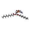


| #8: Chemical |  Mass: 486.726 Da / Num. of mol.: 2 / Source method: obtained synthetically / Formula: C31H50O4 Mass: 486.726 Da / Num. of mol.: 2 / Source method: obtained synthetically / Formula: C31H50O4#9: Chemical |  Mass: 495.587 Da / Num. of mol.: 2 / Source method: obtained synthetically / Formula: C23H46NO8P Mass: 495.587 Da / Num. of mol.: 2 / Source method: obtained synthetically / Formula: C23H46NO8P#10: Chemical |  Mass: 198.388 Da / Num. of mol.: 3 / Source method: obtained synthetically / Formula: C14H30 Mass: 198.388 Da / Num. of mol.: 3 / Source method: obtained synthetically / Formula: C14H30#11: Chemical |  Mass: 170.335 Da / Num. of mol.: 3 / Source method: obtained synthetically / Formula: C12H26 Mass: 170.335 Da / Num. of mol.: 3 / Source method: obtained synthetically / Formula: C12H26#12: Chemical | ChemComp-D10 / |  Mass: 142.282 Da / Num. of mol.: 1 / Source method: obtained synthetically / Formula: C10H22 Mass: 142.282 Da / Num. of mol.: 1 / Source method: obtained synthetically / Formula: C10H22#13: Chemical | ChemComp-A1EOT / [( | Mass: 888.028 Da / Num. of mol.: 1 / Source method: obtained synthetically / Formula: C41H78NO17P / Feature type: SUBJECT OF INVESTIGATION |
|---|
-Details
| Has ligand of interest | Y |
|---|---|
| Has protein modification | Y |
-Experimental details
-Experiment
| Experiment | Method: ELECTRON MICROSCOPY |
|---|---|
| EM experiment | Aggregation state: PARTICLE / 3D reconstruction method: single particle reconstruction |
- Sample preparation
| Component | Name: monomeric glycosylphosphatidylinositol (GPI) transamidase Type: COMPLEX / Entity ID: #1-#5 / Source: RECOMBINANT |
|---|---|
| Molecular weight | Experimental value: NO |
| Source (natural) | Organism: |
| Source (recombinant) | Organism: Homo sapiens (human) |
| Buffer solution | pH: 7.5 |
| Specimen | Embedding applied: NO / Shadowing applied: NO / Staining applied: NO / Vitrification applied: YES |
| Vitrification | Cryogen name: ETHANE |
- Electron microscopy imaging
| Experimental equipment |  Model: Titan Krios / Image courtesy: FEI Company |
|---|---|
| Microscopy | Model: TFS KRIOS |
| Electron gun | Electron source:  FIELD EMISSION GUN / Accelerating voltage: 300 kV / Illumination mode: FLOOD BEAM FIELD EMISSION GUN / Accelerating voltage: 300 kV / Illumination mode: FLOOD BEAM |
| Electron lens | Mode: BRIGHT FIELD / Nominal defocus max: 3000 nm / Nominal defocus min: 1100 nm |
| Image recording | Electron dose: 50 e/Å2 / Film or detector model: GATAN K3 (6k x 4k) |
- Processing
| EM software | Name: PHENIX / Version: 1.20.1_4487: / Category: model refinement |
|---|---|
| CTF correction | Type: PHASE FLIPPING AND AMPLITUDE CORRECTION |
| 3D reconstruction | Resolution: 3.61 Å / Resolution method: FSC 0.143 CUT-OFF / Num. of particles: 55376 / Symmetry type: POINT |
