 Movie
Movie Controller

[English] 日本語
 Yorodumi
- PDB-9u3w: Crystal structure of the ribokinase RBK1 in complex with ribose f... -
+ Open data

- Basic information
| Entry | Database: PDB / ID: 9u3w | ||||||
|---|---|---|---|---|---|---|---|
| Title | Crystal structure of the ribokinase RBK1 in complex with ribose from Saccharomyces cerevisiae | ||||||
 Components Components | Ribokinase | ||||||
 Keywords Keywords | TRANSFERASE / ribokinase | ||||||
| Function / homology |  Function and homology information Function and homology informationribokinase / ribokinase activity / D-ribose catabolic process / ATP binding / metal ion binding / nucleus / cytoplasm / cytosol Similarity search - Function | ||||||
| Biological species |  | ||||||
| Method |  X-RAY DIFFRACTION / SYNCHROTRON / MOLECULAR REPLACEMENT / Resolution: 2.91 Å X-RAY DIFFRACTION / SYNCHROTRON / MOLECULAR REPLACEMENT / Resolution: 2.91 Å | ||||||
 Authors Authors | Yang, X.Y. / Liu, X.H. | ||||||
| Funding support |  China, 1items China, 1items
| ||||||
 Citation Citation | Journal: Int.J.Biol.Macromol. / Year: 2025 Title: Structural and biochemical insights into the molecular mechanism of ribokinase RBK1 from Saccharomyces cerevisiae. Authors: Zhen, S. / Zhang, Z. / Fan, Y. / Li, Y. / Liu, C. / Guo, F. / Zhu, Y. / Wang, Y. / Zhang, J. / Xie, J. / Zhou, H. / Yang, X. / Liu, X. | ||||||
| History |
|
- Structure visualization
| Structure viewer | Molecule: MolmilJmol/JSmol |
|---|
- Downloads & links
-Download
| PDBx/mmCIF format | 9u3w.cif.gz | 130.6 KB | Display | PDBx/mmCIF format |
|---|---|---|---|---|
| PDB format | pdb9u3w.ent.gz | 99.8 KB | Display | PDB format |
| PDBx/mmJSON format | 9u3w.json.gz | Tree view | PDBx/mmJSON format | |
| Others |  Other downloads Other downloads |
-Validation report
| Arichive directory | https://data.pdbj.org/pub/pdb/validation_reports/u3/9u3wftp://data.pdbj.org/pub/pdb/validation_reports/u3/9u3w | HTTPS FTP |
|---|
-Related structure data

-Links
 PDBj
PDBj- Assembly
| Deposited unit | 
| ||||||||
|---|---|---|---|---|---|---|---|---|---|
| 1 | 
| ||||||||
| Unit cell |
|
-Components
| #1: Protein | Mass: 36964.930 Da / Num. of mol.: 2 Source method: isolated from a genetically manipulated source Source: (gene. exp.)  #2: Sugar | 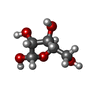  Type: D-saccharide, alpha linking / Mass: 150.130 Da / Num. of mol.: 2 / Source method: obtained synthetically / Formula: C5H10O5 / Feature type: SUBJECT OF INVESTIGATION Type: D-saccharide, alpha linking / Mass: 150.130 Da / Num. of mol.: 2 / Source method: obtained synthetically / Formula: C5H10O5 / Feature type: SUBJECT OF INVESTIGATIONHas ligand of interest | Y | Has protein modification | N | |
|---|
-Experimental details
-Experiment
| Experiment | Method: X-RAY DIFFRACTION / Number of used crystals: 1 |
|---|
- Sample preparation
| Crystal | Density Matthews: 2.72 Å3/Da / Density % sol: 54.84 % |
|---|---|
| Crystal grow | Temperature: 291 K / Method: vapor diffusion Details: 0.2M Sodium formate, 0.1M Bicine pH 8.6, 24% PEG 5000, 0.04% 1,2-Diaminocyclohexane sulfate, 0.04% Diloxanide furoate, 0.04% Fumaric acid, 0.04% Spermine, 0.04% Sulfaguanidine, 0.004M HEPES sodium pH 6.8 |
-Data collection
| Diffraction | Mean temperature: 100 K / Serial crystal experiment: N |
|---|---|
| Diffraction source | Source: SYNCHROTRON / Site: SSRF / Beamline: BL10U2 / Wavelength: 0.978797 Å |
| Detector | Type: DECTRIS EIGER X 16M / Detector: PIXEL / Date: Sep 13, 2023 |
| Radiation | Protocol: SINGLE WAVELENGTH / Monochromatic (M) / Laue (L): M / Scattering type: x-ray |
| Radiation wavelength | Wavelength: 0.978797 Å / Relative weight: 1 |
| Reflection | Resolution: 2.91→84.53 Å / Num. obs: 17881 / % possible obs: 99.2 % / Redundancy: 12.3 % / CC1/2: 0.997 / Net I/σ(I): 14.3 |
| Reflection shell | Resolution: 2.91→2.99 Å / Num. unique obs: 1303 / CC1/2: 0.557 |
- Processing
| Software |
| |||||||||||||||||||||||||||||||||||||||||||||||||
|---|---|---|---|---|---|---|---|---|---|---|---|---|---|---|---|---|---|---|---|---|---|---|---|---|---|---|---|---|---|---|---|---|---|---|---|---|---|---|---|---|---|---|---|---|---|---|---|---|---|---|
| Refinement | Method to determine structure: MOLECULAR REPLACEMENT / Resolution: 2.91→36.47 Å / SU ML: 0.57 / Cross valid method: FREE R-VALUE / σ(F): 1.34 / Phase error: 34.37 / Stereochemistry target values: ML
| |||||||||||||||||||||||||||||||||||||||||||||||||
| Solvent computation | Shrinkage radii: 0.9 Å / VDW probe radii: 1.1 Å / Solvent model: FLAT BULK SOLVENT MODEL | |||||||||||||||||||||||||||||||||||||||||||||||||
| Refinement step | Cycle: LAST / Resolution: 2.91→36.47 Å
| |||||||||||||||||||||||||||||||||||||||||||||||||
| Refine LS restraints |
| |||||||||||||||||||||||||||||||||||||||||||||||||
| LS refinement shell |
|
