Movie
 Controller


+ Open data

- Basic information
| Entry | Database: PDB / ID: 9tzf | |||||||||
|---|---|---|---|---|---|---|---|---|---|---|
| Title | Vibrio cholerae DnaB-DciA open ring complex | |||||||||
 Components |
| |||||||||
 Keywords | REPLICATION / DNA replication initiation / DnaB / replicative helicase / helicase loader / DciA / Vibrio cholerae | |||||||||
| Function / homology | Protein of unknown function DUF721/UPF0232 / Dna[CI] antecedent, DciA / ADENOSINE-5'-DIPHOSPHATE / DciA-like domain-containing protein / :  Function and homology information | |||||||||
| Biological species |  Vibrio cholerae O1 biovar El Tor str. N16961 (bacteria) | |||||||||
| Method | ELECTRON MICROSCOPY / single particle reconstruction / cryo EM / Resolution: 3.5 Å | |||||||||
 Authors | Araujo-Bazan, L. / Arias-Palomo, E. | |||||||||
| Funding support |  Spain, 1items
| |||||||||
 Citation | Journal: Nucleic Acids Res / Year: 2026 Title: Structural basis of DnaB ring opening by the ancestral helicase loader DciA. Authors: Lidia Araújo-Bazán / Ernesto Arias-Palomo / Abstract: Ring-shaped helicases must be opened and loaded onto DNA to initiate genome replication. In bacteria, most species lack canonical AAA+ helicase loaders and instead rely on DciA, an evolutionarily ...Ring-shaped helicases must be opened and loaded onto DNA to initiate genome replication. In bacteria, most species lack canonical AAA+ helicase loaders and instead rely on DciA, an evolutionarily conserved, ATP-independent factor whose mechanism has remained unclear. Here we define how DciA remodels the bacterial replicative helicase DnaB. A 3.5 Å cryo-EM structure reveals a DnaB6·DciA3 complex in which DciA binds the periphery of the helicase and stabilizes a right-handed open-helical conformation with a single cracked interface. Three DciA molecules dock onto alternate linker-helix/docking-helix modules, while one N-terminal domain (NTD) engages the cracked interface, occupying the pocket vacated by the displaced linker helix and acting as a wedge to promote ring opening. Functional analyses reveal how DciA couples ATPase suppression with efficient helicase loading through coordinated actions of its NTDs and C-terminal domains. Together, these findings define a minimal, ATP-independent mechanism for helicase loading and show how an ancestral loader remodels DnaB to overcome a fundamental topological barrier. | |||||||||
| History |
|
- Structure visualization
| Structure viewer | Molecule: MolmilJmol/JSmol |
|---|
- Downloads & links
-Download
| PDBx/mmCIF format | 9tzf.cif.gz | 917.6 KB | Display | PDBx/mmCIF format |
|---|---|---|---|---|
| PDB format | pdb9tzf.ent.gz | 774.3 KB | Display | PDB format |
| PDBx/mmJSON format | 9tzf.json.gz | Tree view | PDBx/mmJSON format | |
| Others |  Other downloads |
-Validation report
| Arichive directory | https://data.pdbj.org/pub/pdb/validation_reports/tz/9tzfftp://data.pdbj.org/pub/pdb/validation_reports/tz/9tzf | HTTPS FTP |
|---|
-Related structure data
| Related structure data |  56467MC M: map data used to model this data C: citing same article ( |
|---|---|
| Similar structure data |
-Links
 PDBj
- Assembly
| Deposited unit | 
|
|---|---|
| 1 |
|
-Components
| #1: Protein | Mass: 52108.773 Da / Num. of mol.: 6 Source method: isolated from a genetically manipulated source Source: (gene. exp.) Vibrio cholerae O1 biovar El Tor str. N16961 (bacteria)Gene: VC_0371 / Plasmid: 2-Cc-T Details (production host): TEV cleavage MBP-His6 tag at the C-terminus Production host:  Escherichia coli (E. coli) / Variant (production host): C41 / References: UniProt: Q9KUY7, DNA 5'-3' helicase #2: Protein | Mass: 17574.307 Da / Num. of mol.: 3 Source method: isolated from a genetically manipulated source Source: (gene. exp.) Vibrio cholerae O1 biovar El Tor str. N16961 (bacteria)Gene: VC_2395 / Plasmid: 1C Details (production host): TEV cleavage MBP-His6 tag at the N-terminus Production host: Escherichia coli BL21(DE3) (bacteria) / References: UniProt: Q9KPH3#3: Chemical | ChemComp-ADP / 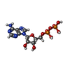  Mass: 427.201 Da / Num. of mol.: 6 Source method: isolated from a genetically manipulated source Formula: C10H15N5O10P2 / Feature type: SUBJECT OF INVESTIGATION / Comment: ADP, energy-carrying molecule*YM #4: Chemical | ChemComp-MG /   Mass: 24.305 Da / Num. of mol.: 6 / Source method: obtained synthetically / Formula: Mg / Feature type: SUBJECT OF INVESTIGATION Has ligand of interest | Y | Has protein modification | N | |
|---|
-Experimental details
-Experiment
| Experiment | Method: ELECTRON MICROSCOPY |
|---|---|
| EM experiment | Aggregation state: PARTICLE / 3D reconstruction method: single particle reconstruction |
- Sample preparation
| Component | Name: VcDnaB-DciA open ring complex / Type: COMPLEX Details: Hexameric DnaB helicase bound to three DciA protomers Entity ID: #1-#2 / Source: RECOMBINANT | ||||||||||||||||||||||||||||||||||||||||
|---|---|---|---|---|---|---|---|---|---|---|---|---|---|---|---|---|---|---|---|---|---|---|---|---|---|---|---|---|---|---|---|---|---|---|---|---|---|---|---|---|---|
| Molecular weight | Value: 0.35 MDa / Experimental value: NO | ||||||||||||||||||||||||||||||||||||||||
| Source (natural) | Organism: Vibrio cholerae O1 biovar El Tor str. N16961 (bacteria) | ||||||||||||||||||||||||||||||||||||||||
| Source (recombinant) | Organism: Escherichia coli (E. coli) / Strain: C41 and BL21 / Plasmid: 2-Cc-T and 1C vectors | ||||||||||||||||||||||||||||||||||||||||
| Buffer solution | pH: 7.5 | ||||||||||||||||||||||||||||||||||||||||
| Buffer component |
| ||||||||||||||||||||||||||||||||||||||||
| Specimen | Embedding applied: NO / Shadowing applied: NO / Staining applied: NO / Vitrification applied: YES | ||||||||||||||||||||||||||||||||||||||||
| Specimen support | Grid material: GOLD / Grid mesh size: 300 divisions/in. / Grid type: Quantifoil R2/1 | ||||||||||||||||||||||||||||||||||||||||
| Vitrification | Instrument: FEI VITROBOT MARK IV / Cryogen name: ETHANE / Humidity: 100 % / Chamber temperature: 298 K |
- Electron microscopy imaging
| Experimental equipment | 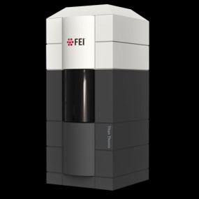 Model: Titan Krios / Image courtesy: FEI Company |
|---|---|
| Microscopy | Model: TFS KRIOS |
| Electron gun | Electron source:  FIELD EMISSION GUN / Accelerating voltage: 300 kV / Illumination mode: FLOOD BEAM |
| Electron lens | Mode: BRIGHT FIELD / Nominal magnification: 105000 X / Nominal defocus max: 2400 nm / Nominal defocus min: 1200 nm / Cs: 2.7 mm |
| Specimen holder | Cryogen: NITROGEN / Specimen holder model: FEI TITAN KRIOS AUTOGRID HOLDER |
| Image recording | Average exposure time: 3.9 sec. / Electron dose: 54.6 e/Å2 / Film or detector model: GATAN K3 BIOQUANTUM (6k x 4k) / Num. of grids imaged: 1 / Num. of real images: 4507 |
- Processing
| EM software |
| ||||||||||||||||||||||||||||||||||||||||
|---|---|---|---|---|---|---|---|---|---|---|---|---|---|---|---|---|---|---|---|---|---|---|---|---|---|---|---|---|---|---|---|---|---|---|---|---|---|---|---|---|---|
| CTF correction | Type: PHASE FLIPPING AND AMPLITUDE CORRECTION | ||||||||||||||||||||||||||||||||||||||||
| Particle selection | Num. of particles selected: 657747 | ||||||||||||||||||||||||||||||||||||||||
| Symmetry | Point symmetry: C1 (asymmetric) | ||||||||||||||||||||||||||||||||||||||||
| 3D reconstruction | Resolution: 3.5 Å / Resolution method: FSC 0.143 CUT-OFF / Num. of particles: 115187 / Num. of class averages: 1 / Symmetry type: POINT | ||||||||||||||||||||||||||||||||||||||||
| Atomic model building | B value: 83.473 / Protocol: OTHER / Space: REAL | ||||||||||||||||||||||||||||||||||||||||
| Refinement | Highest resolution: 3.5 Å Stereochemistry target values: REAL-SPACE (WEIGHTED MAP SUM AT ATOM CENTERS) | ||||||||||||||||||||||||||||||||||||||||
| Refine LS restraints |
|