 Movie
Movie Controller

[English] 日本語
 Yorodumi
- PDB-9ty2: Crystal structure of a Picomolar Nanobody in complex with Maltose... -
+ Open data

- Basic information
| Entry | Database: PDB / ID: 9ty2 | |||||||||
|---|---|---|---|---|---|---|---|---|---|---|
| Title | Crystal structure of a Picomolar Nanobody in complex with Maltose Binding Protein, MBP, and maltose | |||||||||
 Components Components |
| |||||||||
 Keywords Keywords | IMMUNE SYSTEM / Nanobody / Single Chain Antibody / Maltose Binding protein | |||||||||
| Function / homology |  Function and homology information Function and homology informationcarbohydrate transmembrane transporter activity / maltose binding / maltose transport / maltodextrin transmembrane transport / ATP-binding cassette (ABC) transporter complex, substrate-binding subunit-containing / outer membrane-bounded periplasmic space Similarity search - Function | |||||||||
| Biological species |   Escherichia coli (E. coli) Escherichia coli (E. coli)  Vicugna pacos (alpaca) Vicugna pacos (alpaca) | |||||||||
| Method |  X-RAY DIFFRACTION / SYNCHROTRON / MOLECULAR REPLACEMENT / Resolution: 1.75 Å X-RAY DIFFRACTION / SYNCHROTRON / MOLECULAR REPLACEMENT / Resolution: 1.75 Å | |||||||||
 Authors Authors | Chinellato, M. / Franzin, E. / Vascon, F. / Schmit, F. / Pontisso, P. / Cendron, L. | |||||||||
| Funding support |  Italy, 2items Italy, 2items
| |||||||||
 Citation Citation | Journal: Acs Bio Med Chem Au / Year: 2026 Title: A Picomolar MBP-Binding Nanobody for Target Enrichment and Modular Complex Engineering Authors: Chinellato, M. / Franzin, E. / Vascon, F. / Koenig, P.A. / Schmidt, F.I. / Pontisso, P. / Cendron, L. | |||||||||
| History |
|
- Structure visualization
| Structure viewer | Molecule: MolmilJmol/JSmol |
|---|
- Downloads & links
-Download
| PDBx/mmCIF format | 9ty2.cif.gz | 265 KB | Display | PDBx/mmCIF format |
|---|---|---|---|---|
| PDB format | pdb9ty2.ent.gz | 173 KB | Display | PDB format |
| PDBx/mmJSON format | 9ty2.json.gz | Tree view | PDBx/mmJSON format | |
| Others |  Other downloads Other downloads |
-Validation report
| Arichive directory | https://data.pdbj.org/pub/pdb/validation_reports/ty/9ty2ftp://data.pdbj.org/pub/pdb/validation_reports/ty/9ty2 | HTTPS FTP |
|---|
-Related structure data
| Similar structure data | |
|---|---|
| Experimental dataset #1 | Data reference: 10.15151/ESRF-ES-2114514663 / Data set type: diffraction image data |
-Links
 PDBj
PDBj




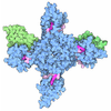
- Assembly
| Deposited unit | 
| ||||||||
|---|---|---|---|---|---|---|---|---|---|
| 1 |
| ||||||||
| Unit cell |
|
-Components
| #1: Protein | Mass: 42554.848 Da / Num. of mol.: 1 Source method: isolated from a genetically manipulated source Details: the missing residues derive from the recombinant construct and are not visible in the electron density maps Source: (gene. exp.) Escherichia coli (E. coli) / Gene: malE, Z5632, ECs5017 / Production host: Escherichia coli (E. coli) / References: UniProt: P0AEY0 |
|---|---|
| #2: Antibody | Mass: 13997.679 Da / Num. of mol.: 1 Source method: isolated from a genetically manipulated source Details: missing residues are not visible in the electron density maps Source: (gene. exp.) Vicugna pacos (alpaca) / Production host:  Komagataella pastoris (fungus) Komagataella pastoris (fungus) |
| #3: Polysaccharide | alpha-D-glucopyranose-(1-4)-beta-D-glucopyranose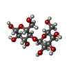 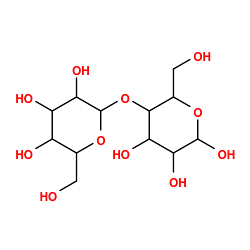 Source method: isolated from a genetically manipulated source Details: oligosaccharide / References: beta-maltose |
| #4: Water | ChemComp-HOH /  Mass: 18.015 Da / Num. of mol.: 372 / Source method: isolated from a natural source / Formula: H2O Mass: 18.015 Da / Num. of mol.: 372 / Source method: isolated from a natural source / Formula: H2O |
| Has ligand of interest | Y |
| Has protein modification | Y |
-Experimental details
-Experiment
| Experiment | Method: X-RAY DIFFRACTION / Number of used crystals: 1 |
|---|
- Sample preparation
| Crystal | Density Matthews: 2.44 Å3/Da / Density % sol: 49.57 % |
|---|---|
| Crystal grow | Temperature: 293 K / Method: vapor diffusion, sitting drop / pH: 7 Details: 0.1 M MIB (Sodium malonate dibasic monohydrate, Imidazole, Boric acid) pH 7.0, 25 % w/v PEG 1500 |
-Data collection
| Diffraction | Mean temperature: 100 K / Serial crystal experiment: N |
|---|---|
| Diffraction source | Source: SYNCHROTRON / Site: ESRF  / Beamline: MASSIF-3 / Wavelength: 0.9677 Å / Beamline: MASSIF-3 / Wavelength: 0.9677 Å |
| Detector | Type: DECTRIS EIGER R 4M / Detector: PIXEL / Date: Mar 7, 2023 |
| Radiation | Protocol: SINGLE WAVELENGTH / Monochromatic (M) / Laue (L): M / Scattering type: x-ray |
| Radiation wavelength | Wavelength: 0.9677 Å / Relative weight: 1 |
| Reflection | Resolution: 1.75→52.23 Å / Num. obs: 54091 / % possible obs: 98 % / Redundancy: 3.8 % / CC1/2: 0.99 / Rmerge(I) obs: 0.164 / Net I/σ(I): 5.5 |
| Reflection shell | Resolution: 1.75→1.78 Å / Rmerge(I) obs: 1.16 / Num. unique obs: 2962 / CC1/2: 0.4 |
- Processing
| Software |
| |||||||||||||||||||||||||||||||||||||||||||||||||||||||||||||||||||||||||||
|---|---|---|---|---|---|---|---|---|---|---|---|---|---|---|---|---|---|---|---|---|---|---|---|---|---|---|---|---|---|---|---|---|---|---|---|---|---|---|---|---|---|---|---|---|---|---|---|---|---|---|---|---|---|---|---|---|---|---|---|---|---|---|---|---|---|---|---|---|---|---|---|---|---|---|---|---|
| Refinement | Method to determine structure: MOLECULAR REPLACEMENT / Resolution: 1.75→52.177 Å / Cor.coef. Fo:Fc: 0.964 / Cor.coef. Fo:Fc free: 0.939 / SU B: 3.807 / SU ML: 0.057 / Cross valid method: FREE R-VALUE / ESU R: 0.038 / ESU R Free: 0.026 / Details: Hydrogens have not been used
| |||||||||||||||||||||||||||||||||||||||||||||||||||||||||||||||||||||||||||
| Solvent computation | Ion probe radii: 0.8 Å / Shrinkage radii: 0.8 Å / VDW probe radii: 1.2 Å / Solvent model: MASK BULK SOLVENT | |||||||||||||||||||||||||||||||||||||||||||||||||||||||||||||||||||||||||||
| Displacement parameters | Biso mean: 26.013 Å2
| |||||||||||||||||||||||||||||||||||||||||||||||||||||||||||||||||||||||||||
| Refinement step | Cycle: LAST / Resolution: 1.75→52.177 Å
| |||||||||||||||||||||||||||||||||||||||||||||||||||||||||||||||||||||||||||
| LS refinement shell | Resolution: 1.75→1.795 Å
| |||||||||||||||||||||||||||||||||||||||||||||||||||||||||||||||||||||||||||
| Refinement TLS params. | Method: refined / Refine-ID: X-RAY DIFFRACTION
| |||||||||||||||||||||||||||||||||||||||||||||||||||||||||||||||||||||||||||
| Refinement TLS group | Selection: ALL |
