oxidoreductase activity, acting on CH or CH2 groups, with an iron-sulfur protein as acceptor / 4 iron, 4 sulfur cluster binding / heme binding / metal ion binding Similarity search - Function
Mass: 18.015 Da / Num. of mol.: 789 / Source method: isolated from a natural source / Formula: H2O
-
Details
Has ligand of interest
Y
Has protein modification
Y
-
Experimental details
-
Experiment
Experiment
Method: X-RAY DIFFRACTION / Number of used crystals: 1
-
Sample preparation
Crystal
Density Matthews: 2.8 Å3/Da / Density % sol: 56.17 % / Description: Dark brown trapezoidal thick plate
Crystal grow
Temperature: 293.15 K / Method: vapor diffusion, sitting drop / pH: 4.6 Details: Prior to crystallisation, fresh sample was centrifuged at 13 000 g for 3 min to remove macro-aggregates and dust. The sample was crystallised in an anaerobic chamber filled with a N2/H2 (97: ...Details: Prior to crystallisation, fresh sample was centrifuged at 13 000 g for 3 min to remove macro-aggregates and dust. The sample was crystallised in an anaerobic chamber filled with a N2/H2 (97:3%) atmosphere, at 20 degrees Celsius. The crystallisation was done in 96-Well MRC 2-Drop polystyrene plates (SWISSCI) containing 90 uL of crystallisation solution in the reservoir. 0.7 uL of enzyme at 6.5 mg/ml + 1 mM FAD was mixed with 0.7 uL of the crystallisation solution. The crystallisation solution contained 45 % w/v pentaerythritol propoxylate (5/4 PO/OH), 100 mM sodium acetate pH 4.6, and 400 mM potassium chloride. Prior to flash-freezing in liquid nitrogen, crystals were soaked in the crystallisation solution supplemented with 50 mM sodium nitrite for 5 min.
-
Data collection
Diffraction
Mean temperature: 100 K / Serial crystal experiment: N
Method to determine structure: MOLECULAR REPLACEMENT / Resolution: 1.2→42.02 Å / SU ML: 0.08 / Cross valid method: FREE R-VALUE / σ(F): 1.34 / Phase error: 15.49 / Stereochemistry target values: ML Details: The refinement has been performed with phenix.refine considering all atoms anisotropic. The refinement has been done with hydrogens in riding position.
Rfactor
Num. reflection
% reflection
Rfree
0.1427
8740
4.99 %
Rwork
0.1157
-
-
obs
0.117
175289
73.2 %
Solvent computation
Shrinkage radii: 0.9 Å / VDW probe radii: 1.1 Å / Solvent model: FLAT BULK SOLVENT MODEL
Displacement parameters
Biso mean: 20.51 Å2
Refinement step
Cycle: LAST / Resolution: 1.2→42.02 Å
Protein
Nucleic acid
Ligand
Solvent
Total
Num. atoms
4894
0
222
789
5905
Refine LS restraints
Refine-ID
Type
Dev ideal
Number
X-RAY DIFFRACTION
f_bond_d
0.013
5384
X-RAY DIFFRACTION
f_angle_d
1.481
7296
X-RAY DIFFRACTION
f_dihedral_angle_d
15.814
2147
X-RAY DIFFRACTION
f_chiral_restr
0.106
778
X-RAY DIFFRACTION
f_plane_restr
0.017
900
LS refinement shell
Resolution (Å)
Rfactor Rfree
Num. reflection Rfree
Rfactor Rwork
Num. reflection Rwork
Refine-ID
% reflection obs (%)
1.2-1.22
0.1124
2
0.2387
76
X-RAY DIFFRACTION
1
1.22-1.23
0.2894
14
0.2527
259
X-RAY DIFFRACTION
3
1.23-1.25
0.2284
29
0.2528
490
X-RAY DIFFRACTION
7
1.25-1.26
0.2578
47
0.2454
712
X-RAY DIFFRACTION
10
1.26-1.28
0.2477
63
0.2392
1084
X-RAY DIFFRACTION
14
1.28-1.3
0.2648
72
0.2412
1630
X-RAY DIFFRACTION
22
1.3-1.32
0.2562
137
0.2279
2361
X-RAY DIFFRACTION
32
1.32-1.34
0.2465
197
0.2231
3549
X-RAY DIFFRACTION
47
1.34-1.36
0.245
281
0.2143
4600
X-RAY DIFFRACTION
61
1.36-1.38
0.2332
294
0.1938
5197
X-RAY DIFFRACTION
69
1.38-1.4
0.2004
289
0.1872
5649
X-RAY DIFFRACTION
75
1.4-1.43
0.2097
322
0.1743
6106
X-RAY DIFFRACTION
81
1.43-1.46
0.1968
314
0.1612
6757
X-RAY DIFFRACTION
89
1.46-1.49
0.1952
348
0.1535
7026
X-RAY DIFFRACTION
93
1.49-1.52
0.1742
390
0.146
7286
X-RAY DIFFRACTION
96
1.52-1.55
0.1532
385
0.1315
7449
X-RAY DIFFRACTION
99
1.55-1.59
0.152
395
0.1264
7586
X-RAY DIFFRACTION
100
1.59-1.63
0.1383
392
0.1156
7549
X-RAY DIFFRACTION
100
1.63-1.68
0.1388
391
0.1032
7561
X-RAY DIFFRACTION
100
1.68-1.74
0.1242
376
0.0989
7595
X-RAY DIFFRACTION
100
1.74-1.8
0.131
426
0.0968
7520
X-RAY DIFFRACTION
100
1.8-1.87
0.1144
375
0.0967
7587
X-RAY DIFFRACTION
100
1.87-1.96
0.1152
411
0.0939
7599
X-RAY DIFFRACTION
100
1.96-2.06
0.1251
396
0.0933
7526
X-RAY DIFFRACTION
99
2.06-2.19
0.1255
419
0.0926
7546
X-RAY DIFFRACTION
100
2.19-2.36
0.1205
410
0.0954
7598
X-RAY DIFFRACTION
99
2.36-2.59
0.1307
376
0.1024
7606
X-RAY DIFFRACTION
99
2.59-2.97
0.1331
391
0.1134
7594
X-RAY DIFFRACTION
99
2.97-3.74
0.1421
405
0.1158
7622
X-RAY DIFFRACTION
99
3.74-42.02
0.1509
393
0.124
7829
X-RAY DIFFRACTION
98
+
About Yorodumi
-
News
-
Feb 9, 2022. New format data for meta-information of EMDB entries
New format data for meta-information of EMDB entries
Version 3 of the EMDB header file is now the official format.
The previous official version 1.9 will be removed from the archive.
In the structure databanks used in Yorodumi, some data are registered as the other names, "COVID-19 virus" and "2019-nCoV". Here are the details of the virus and the list of structure data.
Jan 31, 2019. EMDB accession codes are about to change! (news from PDBe EMDB page)
EMDB accession codes are about to change! (news from PDBe EMDB page)
The allocation of 4 digits for EMDB accession codes will soon come to an end. Whilst these codes will remain in use, new EMDB accession codes will include an additional digit and will expand incrementally as the available range of codes is exhausted. The current 4-digit format prefixed with “EMD-” (i.e. EMD-XXXX) will advance to a 5-digit format (i.e. EMD-XXXXX), and so on. It is currently estimated that the 4-digit codes will be depleted around Spring 2019, at which point the 5-digit format will come into force.
The EM Navigator/Yorodumi systems omit the EMD- prefix.
Related info.:Q: What is EMD? / ID/Accession-code notation in Yorodumi/EM Navigator
Yorodumi is a browser for structure data from EMDB, PDB, SASBDB, etc.
This page is also the successor to EM Navigator detail page, and also detail information page/front-end page for Omokage search.
The word "yorodu" (or yorozu) is an old Japanese word meaning "ten thousand". "mi" (miru) is to see.
Related info.:EMDB / PDB / SASBDB / Comparison of 3 databanks / Yorodumi Search / Aug 31, 2016. New EM Navigator & Yorodumi / Yorodumi Papers / Jmol/JSmol / Function and homology information / Changes in new EM Navigator and Yorodumi
 Movie
Movie Controller

 Yorodumi
 Open data
 Basic information
 Components
 Keywords
 Function and homology information
 Methanocaldococcus infernus ME (archaea)
 X-RAY DIFFRACTION /
  Authors
 Germany,
  France, 2items
  Citation
 Structure visualization
 Downloads & links
 Other downloads
 PDBj
PDBj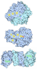
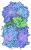
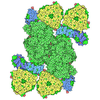
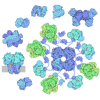
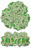


 Assembly

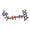




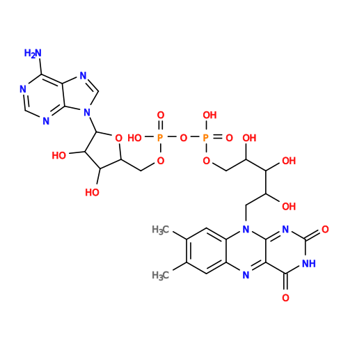 Mass: 785.550 Da / Num. of mol.: 1 / Source method: obtained synthetically / Formula: C27H33N9O15P2 / Feature type: SUBJECT OF INVESTIGATION / Comment: FAD*YM
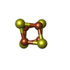
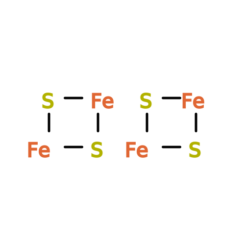 Mass: 351.640 Da / Num. of mol.: 6 / Source method: obtained synthetically / Formula: Fe4S4 / Feature type: SUBJECT OF INVESTIGATION
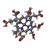
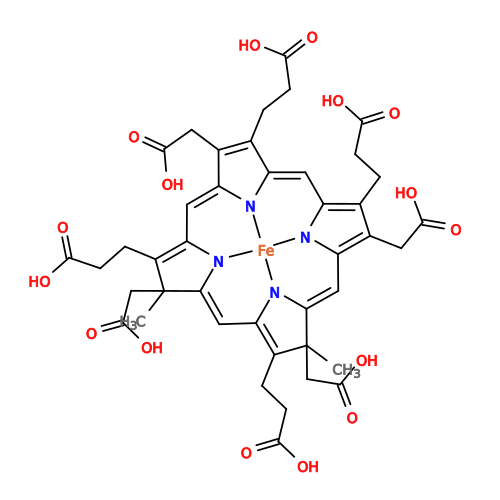 Mass: 916.661 Da / Num. of mol.: 1 / Source method: obtained synthetically / Formula: C42H44FeN4O16 / Feature type: SUBJECT OF INVESTIGATION
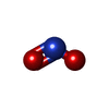
 Mass: 46.005 Da / Num. of mol.: 1 / Source method: obtained synthetically / Formula: NO2 / Feature type: SUBJECT OF INVESTIGATION
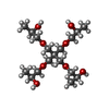
 Mass: 368.463 Da / Num. of mol.: 2 / Source method: obtained synthetically / Formula: C17H36O8
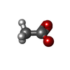
 Mass: 59.044 Da / Num. of mol.: 1 / Source method: obtained synthetically / Formula: C2H3O2
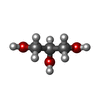
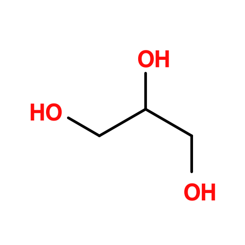 Mass: 92.094 Da / Num. of mol.: 3 / Source method: obtained synthetically / Formula: C3H8O3
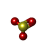
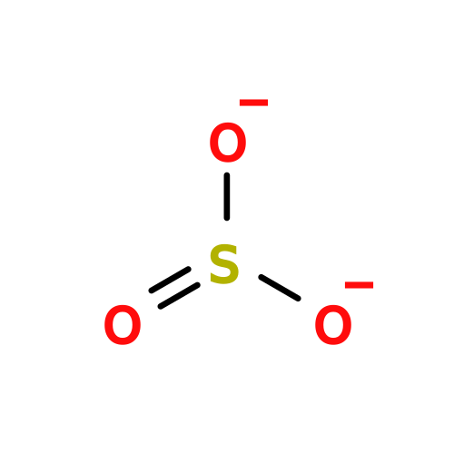 Mass: 80.063 Da / Num. of mol.: 1 / Source method: obtained synthetically / Formula: SO3 / Feature type: SUBJECT OF INVESTIGATION
 Mass: 22.990 Da / Num. of mol.: 1 / Source method: obtained synthetically / Formula: Na
 Mass: 39.098 Da / Num. of mol.: 1 / Source method: obtained synthetically / Formula: K
 Mass: 35.453 Da / Num. of mol.: 2 / Source method: obtained synthetically / Formula: Cl
 Sample preparation
 Processing