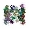
Movie
Movie Controller

[English] 日本語
 Yorodumi
- PDB-9tlk: Cryo-EM Structure of the oligomeric LPOR:Chlide:NADPH Complexes HF-25 -
+ Open data

- Basic information
| Entry | Database: PDB / ID: 9tlk | ||||||||||||||||||||||||
|---|---|---|---|---|---|---|---|---|---|---|---|---|---|---|---|---|---|---|---|---|---|---|---|---|---|
| Title | Cryo-EM Structure of the oligomeric LPOR:Chlide:NADPH Complexes HF-25 | ||||||||||||||||||||||||
 Components Components | Protochlorophyllide reductase B, chloroplastic | ||||||||||||||||||||||||
 Keywords Keywords | PHOTOSYNTHESIS / photoenzyme / chlorophyllide / oligomer | ||||||||||||||||||||||||
| Function / homology |  Function and homology information Function and homology informationprotochlorophyllide reductase / protochlorophyllide reductase activity / response to ethylene / chloroplast outer membrane / chlorophyll biosynthetic process / chloroplast thylakoid / chloroplast envelope / chloroplast thylakoid membrane / photosynthesis / chloroplast ...protochlorophyllide reductase / protochlorophyllide reductase activity / response to ethylene / chloroplast outer membrane / chlorophyll biosynthetic process / chloroplast thylakoid / chloroplast envelope / chloroplast thylakoid membrane / photosynthesis / chloroplast / protein domain specific binding / mRNA binding / cytosol Similarity search - Function | ||||||||||||||||||||||||
| Biological species |   Arabidopsis thaliana (thale cress) Arabidopsis thaliana (thale cress) | ||||||||||||||||||||||||
| Method | ELECTRON MICROSCOPY / helical reconstruction / cryo EM / Resolution: 3.2 Å | ||||||||||||||||||||||||
 Authors Authors | Gabruk, M. / Desfosses, A. / Estrozi, L.F. / Pintscher, S. / Rawski, M. | ||||||||||||||||||||||||
| Funding support |  Poland, 1items Poland, 1items
| ||||||||||||||||||||||||
 Citation Citation | Journal: Nat Commun / Year: 2026 Title: Structures of LPOR-Chlide complexes reveal the structural basis of membrane remodeling and photocatalysis. Authors: Michał Gabruk / Ambroise Desfosses / Leandro Farias Estrozi / Sebastian Pintscher / Michał Rawski / Grzegorz Ważny / Agnieszka Garbacz / Mateusz Zbyradowski / Jerzy Kruk / Leszek Fiedor /  Abstract: Light-dependent protochlorophyllide oxidoreductase (LPOR) is a light-driven enzyme in flowering plants. It is involved in chlorophyll biosynthesis while also reorganizing membrane lipids into the ...Light-dependent protochlorophyllide oxidoreductase (LPOR) is a light-driven enzyme in flowering plants. It is involved in chlorophyll biosynthesis while also reorganizing membrane lipids into the cubic membrane network that supports chloroplast development. However, the structural basis of these two activities and their relationship have remained unclear. Here, cryo-electron microscopy of chlorophyllide-bound LPOR oligomers reveals nine distinct assembly states, including helical filaments, stacked rings and segmented strings of dimers. We find that strings of LPOR dimers reshape lipid bilayers into a range of membrane architectures through combinations of three inter-string interfaces, providing a structural explanation for the flexibility of these assemblies. The highest-resolution map (2.55 Å), shows the pigment-binding region in sufficient detail to reveal a solvent-accessible channel near the pigment and a conformation of the propionate group may support hydride transfer from NADPH. Together, these findings establish a structural framework linking LPOR oligomerization, membrane remodeling and photocatalysis, and suggest that chlorophyllide-bound LPOR assemblies may have a regulatory function in mature leaves. | ||||||||||||||||||||||||
| History |
|
- Structure visualization
| Structure viewer | Molecule: MolmilJmol/JSmol |
|---|
- Downloads & links
-Download
| PDBx/mmCIF format | 9tlk.cif.gz | 5.3 MB | Display | PDBx/mmCIF format |
|---|---|---|---|---|
| PDB format | pdb9tlk.ent.gz | Display | PDB format | |
| PDBx/mmJSON format | 9tlk.json.gz | Tree view | PDBx/mmJSON format | |
| Others |  Other downloads Other downloads |
-Validation report
| Arichive directory | https://data.pdbj.org/pub/pdb/validation_reports/tl/9tlkftp://data.pdbj.org/pub/pdb/validation_reports/tl/9tlk | HTTPS FTP |
|---|
-Related structure data
| Related structure data |  56056MC  9tl6C  9tl7C  9tl8C  9tl9C  9tlaC  9tlbC  9tlcC  9tlhC M: map data used to model this data C: citing same article ( |
|---|---|
| Similar structure data |


-Links
 PDBj
PDBj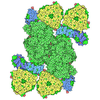
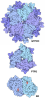
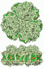

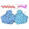
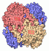
- Assembly
| Deposited unit | 
|
|---|---|
| 1 |
|
-Components
| #1: Protein | Mass: 38236.324 Da / Num. of mol.: 48 Source method: isolated from a genetically manipulated source Source: (gene. exp.) Arabidopsis thaliana (thale cress) / Gene: PORB, At4g27440, F27G19.40 / Plasmid: pET15b / Production host:   Escherichia coli BL21(DE3) (bacteria) / Variant (production host): pRIL / References: UniProt: P21218, protochlorophyllide reductase Escherichia coli BL21(DE3) (bacteria) / Variant (production host): pRIL / References: UniProt: P21218, protochlorophyllide reductase#2: Chemical | ChemComp-NDP / 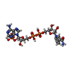 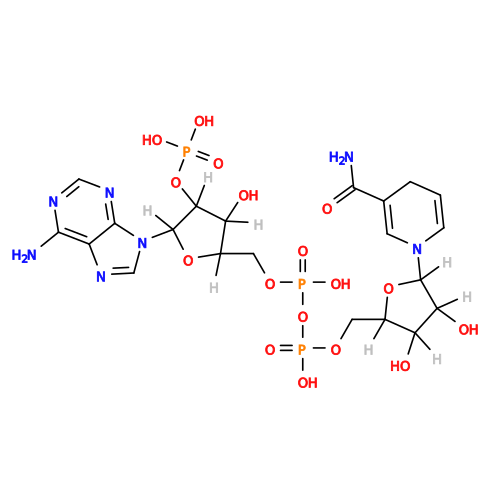 Mass: 745.421 Da / Num. of mol.: 48 / Source method: obtained synthetically / Formula: C21H30N7O17P3 / Feature type: SUBJECT OF INVESTIGATION Mass: 745.421 Da / Num. of mol.: 48 / Source method: obtained synthetically / Formula: C21H30N7O17P3 / Feature type: SUBJECT OF INVESTIGATION#3: Chemical | ChemComp-LMG /  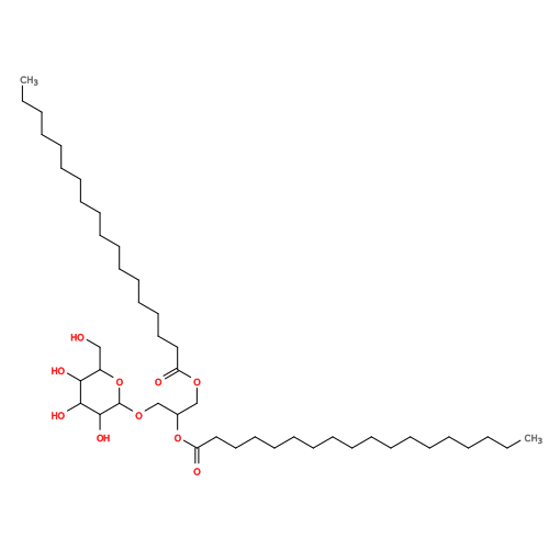 Mass: 787.158 Da / Num. of mol.: 48 / Source method: obtained synthetically / Formula: C45H86O10 / Feature type: SUBJECT OF INVESTIGATION Mass: 787.158 Da / Num. of mol.: 48 / Source method: obtained synthetically / Formula: C45H86O10 / Feature type: SUBJECT OF INVESTIGATION#4: Chemical | ChemComp-A1JWG / Mass: 614.973 Da / Num. of mol.: 48 / Source method: obtained synthetically / Formula: C35H34MgN4O5 / Feature type: SUBJECT OF INVESTIGATION Has ligand of interest | Y | Has protein modification | N | |
|---|
-Experimental details
-Experiment
| Experiment | Method: ELECTRON MICROSCOPY |
|---|---|
| EM experiment | Aggregation state: HELICAL ARRAY / 3D reconstruction method: helical reconstruction |
- Sample preparation
| Component | Name: Cryo-EM Structure of the oligomeric LPOR:Chlide:NADPH Complexes HF-25 Type: COMPLEX / Entity ID: #1 / Source: RECOMBINANT | ||||||||||||||||||||||||||||||
|---|---|---|---|---|---|---|---|---|---|---|---|---|---|---|---|---|---|---|---|---|---|---|---|---|---|---|---|---|---|---|---|
| Molecular weight | Experimental value: NO | ||||||||||||||||||||||||||||||
| Source (natural) | Organism: Arabidopsis thaliana (thale cress) | ||||||||||||||||||||||||||||||
| Source (recombinant) | Organism: Escherichia coli (E. coli) / Strain: BL21(DE3)pRIL / Plasmid: pET15b | ||||||||||||||||||||||||||||||
| Buffer solution | pH: 7.1 | ||||||||||||||||||||||||||||||
| Buffer component |
| ||||||||||||||||||||||||||||||
| Specimen | Embedding applied: NO / Shadowing applied: NO / Staining applied: NO / Vitrification applied: YES Details: 28 uM AtPORB, 40 uM Chlorophyllide, 600 uM NADPH, 230 uM lipids (50mol% MGDG, 35mol% DGDG, 15mol% PG) | ||||||||||||||||||||||||||||||
| Vitrification | Instrument: FEI VITROBOT MARK III / Cryogen name: ETHANE / Humidity: 100 % / Chamber temperature: 294 K |
- Electron microscopy imaging
| Experimental equipment |  Model: Titan Krios / Image courtesy: FEI Company |
|---|---|
| Microscopy | Model: TFS KRIOS |
| Electron gun | Electron source:  FIELD EMISSION GUN / Accelerating voltage: 300 kV / Illumination mode: FLOOD BEAM FIELD EMISSION GUN / Accelerating voltage: 300 kV / Illumination mode: FLOOD BEAM |
| Electron lens | Mode: BRIGHT FIELD / Nominal defocus max: 2100 nm / Nominal defocus min: 900 nm |
| Image recording | Electron dose: 40 e/Å2 / Film or detector model: GATAN K3 BIOQUANTUM (6k x 4k) |
- Processing
| EM software |
| ||||||||||||||||
|---|---|---|---|---|---|---|---|---|---|---|---|---|---|---|---|---|---|
| CTF correction | Type: PHASE FLIPPING AND AMPLITUDE CORRECTION | ||||||||||||||||
| Helical symmerty | Angular rotation/subunit: -114.391 ° / Axial rise/subunit: 25.609 Å / Axial symmetry: D4 | ||||||||||||||||
| 3D reconstruction | Resolution: 3.2 Å / Resolution method: FSC 0.143 CUT-OFF / Num. of particles: 23139 / Symmetry type: HELICAL | ||||||||||||||||
| Atomic model building | PDB-ID: 7JK9 Accession code: 7JK9 / Details: initial model / Source name: PDB / Type: experimental model |