 Movie
Movie Controller

[English] 日本語
 Yorodumi
- PDB-9tbb: Crystal structure of the CsPYL1(I91L-V112L-V192L-L195C-K199S)-ABA... -
+ Open data

- Basic information
| Entry | Database: PDB / ID: 9tbb | ||||||
|---|---|---|---|---|---|---|---|
| Title | Crystal structure of the CsPYL1(I91L-V112L-V192L-L195C-K199S)-ABA-HAB1 ternary complex | ||||||
 Components Components |
| ||||||
 Keywords Keywords | PLANT PROTEIN / ABA receptor / inhibitory ternary complex | ||||||
| Function / homology |  Function and homology information Function and homology informationprotein phosphatase inhibitor complex / regulation of intracellular signal transduction / abscisic acid binding / abscisic acid-activated signaling pathway / protein phosphatase inhibitor activity / protein-serine/threonine phosphatase / protein serine/threonine phosphatase activity / signaling receptor activity / protein homodimerization activity / metal ion binding ...protein phosphatase inhibitor complex / regulation of intracellular signal transduction / abscisic acid binding / abscisic acid-activated signaling pathway / protein phosphatase inhibitor activity / protein-serine/threonine phosphatase / protein serine/threonine phosphatase activity / signaling receptor activity / protein homodimerization activity / metal ion binding / nucleus / cytoplasm Similarity search - Function | ||||||
| Biological species |  | ||||||
| Method |  X-RAY DIFFRACTION / SYNCHROTRON / MOLECULAR REPLACEMENT / Resolution: 1.91 Å X-RAY DIFFRACTION / SYNCHROTRON / MOLECULAR REPLACEMENT / Resolution: 1.91 Å | ||||||
 Authors Authors | Rivera-Moreno, M. / Infantes, L. / Albert, A. | ||||||
| Funding support |  Spain, 1items Spain, 1items
| ||||||
 Citation Citation | Journal: Proc.Natl.Acad.Sci.USA / Year: 2026 Title: Evolutionary-based remodeling of ABA receptors reveals the structural basis of hormone perception and regulation. Authors: Rivera-Moreno, M. / Bono, M. / Infantes, L. / Rodriguez, P.L. / Albert, A. | ||||||
| History |
|
- Structure visualization
| Structure viewer | Molecule: MolmilJmol/JSmol |
|---|
- Downloads & links
-Download
| PDBx/mmCIF format | 9tbb.cif.gz | 140 KB | Display | PDBx/mmCIF format |
|---|---|---|---|---|
| PDB format | pdb9tbb.ent.gz | 85.5 KB | Display | PDB format |
| PDBx/mmJSON format | 9tbb.json.gz | Tree view | PDBx/mmJSON format | |
| Others |  Other downloads Other downloads |
-Validation report
| Arichive directory | https://data.pdbj.org/pub/pdb/validation_reports/tb/9tbbftp://data.pdbj.org/pub/pdb/validation_reports/tb/9tbb | HTTPS FTP |
|---|
-Related structure data
| Related structure data |  9tb5C  9tb6C  9tb7C  9tb8C  9tb9C  9tbaC  9tbcC C: citing same article ( |
|---|---|
| Similar structure data |
-Links
 PDBj
PDBj- Assembly
| Deposited unit | 
| ||||||||||||
|---|---|---|---|---|---|---|---|---|---|---|---|---|---|
| 1 |
| ||||||||||||
| Unit cell |
|
-Components
-Protein , 2 types, 2 molecules AB
| #1: Protein | Mass: 23270.744 Da / Num. of mol.: 1 / Mutation: I91L-V112L-V192L-L195C-K199S Source method: isolated from a genetically manipulated source Source: (gene. exp.)  |
|---|---|
| #2: Protein | Mass: 37029.473 Da / Num. of mol.: 1 Source method: isolated from a genetically manipulated source Source: (gene. exp.) References: UniProt: Q9CAJ0, protein-serine/threonine phosphatase |
-Non-polymers , 4 types, 137 molecules 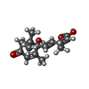


| #3: Chemical | ChemComp-A8S / ( Mass: 264.317 Da / Num. of mol.: 1 / Source method: obtained synthetically / Formula: C15H20O4 / Feature type: SUBJECT OF INVESTIGATION / Comment: hormone*YM Mass: 264.317 Da / Num. of mol.: 1 / Source method: obtained synthetically / Formula: C15H20O4 / Feature type: SUBJECT OF INVESTIGATION / Comment: hormone*YM | ||
|---|---|---|---|
| #4: Chemical | ChemComp-GOL /  Mass: 92.094 Da / Num. of mol.: 1 / Source method: obtained synthetically / Formula: C3H8O3 Mass: 92.094 Da / Num. of mol.: 1 / Source method: obtained synthetically / Formula: C3H8O3 | ||
| #5: Chemical |  Mass: 54.938 Da / Num. of mol.: 3 / Source method: obtained synthetically / Formula: Mn Mass: 54.938 Da / Num. of mol.: 3 / Source method: obtained synthetically / Formula: Mn#6: Water | ChemComp-HOH / | Mass: 18.015 Da / Num. of mol.: 132 / Source method: isolated from a natural source / Formula: H2O |
-Details
| Has ligand of interest | Y |
|---|---|
| Has protein modification | N |
-Experimental details
-Experiment
| Experiment | Method: X-RAY DIFFRACTION / Number of used crystals: 1 |
|---|
- Sample preparation
| Crystal | Density Matthews: 2.17 Å3/Da / Density % sol: 43.3 % |
|---|---|
| Crystal grow | Temperature: 291 K / Method: microbatch / pH: 7 Details: 0.1 M Bis-Tris propane, pH 7.0, 0.5 M CaCl2, 25% (w/v) PEG 3350 |
-Data collection
| Diffraction | Mean temperature: 100 K / Serial crystal experiment: N |
|---|---|
| Diffraction source | Source: SYNCHROTRON / Site: ALBA / Beamline: XALOC / Wavelength: 0.97926 Å |
| Detector | Type: DECTRIS PILATUS 6M / Detector: PIXEL / Date: Sep 10, 2025 |
| Radiation | Monochromator: M / Protocol: SINGLE WAVELENGTH / Monochromatic (M) / Laue (L): M / Scattering type: x-ray |
| Radiation wavelength | Wavelength: 0.97926 Å / Relative weight: 1 |
| Reflection | Resolution: 1.9→46.72 Å / Num. obs: 22383 / % possible obs: 99.8 % / Redundancy: 90.9 % / Biso Wilson estimate: 18.51 Å2 / CC1/2: 0.991 / Net I/σ(I): 7.1 |
| Reflection shell | Resolution: 1.9→1.99 Å / Num. unique obs: 1119 / CC1/2: 0.598 |
- Processing
| Software |
| |||||||||||||||||||||||||||||||||||||||||||||||||||||||||||||||
|---|---|---|---|---|---|---|---|---|---|---|---|---|---|---|---|---|---|---|---|---|---|---|---|---|---|---|---|---|---|---|---|---|---|---|---|---|---|---|---|---|---|---|---|---|---|---|---|---|---|---|---|---|---|---|---|---|---|---|---|---|---|---|---|---|
| Refinement | Method to determine structure: MOLECULAR REPLACEMENT / Resolution: 1.91→46.72 Å / SU ML: 0.2467 / Cross valid method: FREE R-VALUE / σ(F): 1.34 / Phase error: 30.703 Stereochemistry target values: GeoStd + Monomer Library + CDL v1.2
| |||||||||||||||||||||||||||||||||||||||||||||||||||||||||||||||
| Solvent computation | Shrinkage radii: 0.9 Å / VDW probe radii: 1.1 Å / Solvent model: FLAT BULK SOLVENT MODEL | |||||||||||||||||||||||||||||||||||||||||||||||||||||||||||||||
| Displacement parameters | Biso mean: 24.71 Å2 | |||||||||||||||||||||||||||||||||||||||||||||||||||||||||||||||
| Refinement step | Cycle: LAST / Resolution: 1.91→46.72 Å
| |||||||||||||||||||||||||||||||||||||||||||||||||||||||||||||||
| Refine LS restraints |
| |||||||||||||||||||||||||||||||||||||||||||||||||||||||||||||||
| LS refinement shell |
|
