 Movie
Movie Controller

[English] 日本語
 Yorodumi
- PDB-9spz: Crystal structure of the Molybdenum-containing nitrogenase from M... -
+ Open data

- Basic information
| Entry | Database: PDB / ID: 9spz | ||||||
|---|---|---|---|---|---|---|---|
| Title | Crystal structure of the Molybdenum-containing nitrogenase from Methanocaldococcus infernus refined to 1.37 A resolution - crystalline form A | ||||||
 Components Components |
| ||||||
 Keywords Keywords | OXIDOREDUCTASE / Nitrogenase / N2-fixation / FeMo-cofactor / hyperthermophile / extremophile / metallo-cofactor / metalloenzyme / P-cluster / archaea / methanogen / marine / hydrogenotrophic / Methanococcales. | ||||||
| Function / homology |  Function and homology information Function and homology informationnitrogenase / nitrogenase activity / iron-sulfur cluster binding / metal ion binding / ATP binding Similarity search - Function | ||||||
| Biological species |  Methanocaldococcus infernus ME (archaea) Methanocaldococcus infernus ME (archaea) | ||||||
| Method |  X-RAY DIFFRACTION / SYNCHROTRON / MOLECULAR REPLACEMENT / Resolution: 1.37 Å X-RAY DIFFRACTION / SYNCHROTRON / MOLECULAR REPLACEMENT / Resolution: 1.37 Å | ||||||
 Authors Authors | Maslac, N. / Torer, M.R. / Bolte, P. / Wagner, T. | ||||||
| Funding support |  Germany, 1items Germany, 1items
| ||||||
 Citation Citation | Journal: Nat Commun / Year: 2026 Title: Molecular basis of N 2 fixation in a hyperthermophilic archaeon. Authors: Maslac, N. / Torer, M.R. / Bolte, P. / Wagner, T. | ||||||
| History |
|
- Structure visualization
| Structure viewer | Molecule: MolmilJmol/JSmol |
|---|
- Downloads & links
-Download
| PDBx/mmCIF format | 9spz.cif.gz | 1.1 MB | Display | PDBx/mmCIF format |
|---|---|---|---|---|
| PDB format | pdb9spz.ent.gz | 968.4 KB | Display | PDB format |
| PDBx/mmJSON format | 9spz.json.gz | Tree view | PDBx/mmJSON format | |
| Others |  Other downloads Other downloads |
-Validation report
| Arichive directory | https://data.pdbj.org/pub/pdb/validation_reports/sp/9spzftp://data.pdbj.org/pub/pdb/validation_reports/sp/9spz | HTTPS FTP |
|---|
-Related structure data

-Links
 PDBj
PDBj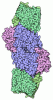
- Assembly
| Deposited unit | 
| ||||||||
|---|---|---|---|---|---|---|---|---|---|
| 1 |
| ||||||||
| Unit cell |
|
-Components
-Protein , 2 types, 4 molecules ACBD
| #1: Protein | Mass: 54738.129 Da / Num. of mol.: 2 / Source method: isolated from a natural source / Source: (natural) Methanocaldococcus infernus ME (archaea) / Cell line: / / Organ: / / Plasmid details: / / Variant: / / Strain: ME / Tissue: / / References: UniProt: D5VU98, nitrogenase#2: Protein | Mass: 51823.508 Da / Num. of mol.: 2 / Source method: isolated from a natural source / Source: (natural) Methanocaldococcus infernus ME (archaea) / Cell line: / / Organ: / / Plasmid details: / / Variant: / / Strain: ME / Tissue: / / References: UniProt: D5VU97, nitrogenase |
|---|
-Non-polymers , 12 types, 1489 molecules 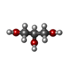
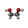

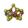




| #3: Chemical | ChemComp-GOL / 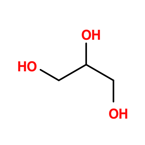 Mass: 92.094 Da / Num. of mol.: 9 / Source method: obtained synthetically / Formula: C3H8O3 Mass: 92.094 Da / Num. of mol.: 9 / Source method: obtained synthetically / Formula: C3H8O3#4: Chemical | ChemComp-EDO / 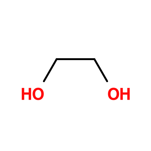 Mass: 62.068 Da / Num. of mol.: 8 / Source method: obtained synthetically / Formula: C2H6O2 Mass: 62.068 Da / Num. of mol.: 8 / Source method: obtained synthetically / Formula: C2H6O2#5: Chemical |  Mass: 671.215 Da / Num. of mol.: 2 / Source method: obtained synthetically / Formula: Fe8S7 / Feature type: SUBJECT OF INVESTIGATION Mass: 671.215 Da / Num. of mol.: 2 / Source method: obtained synthetically / Formula: Fe8S7 / Feature type: SUBJECT OF INVESTIGATION#6: Chemical | 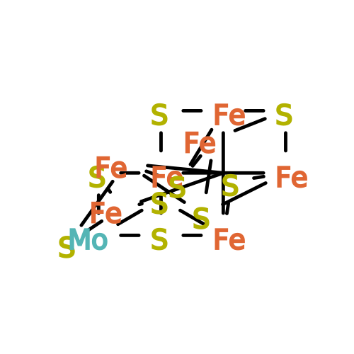 Mass: 787.451 Da / Num. of mol.: 2 / Source method: obtained synthetically / Formula: CFe7MoS9 / Feature type: SUBJECT OF INVESTIGATION Mass: 787.451 Da / Num. of mol.: 2 / Source method: obtained synthetically / Formula: CFe7MoS9 / Feature type: SUBJECT OF INVESTIGATION#7: Chemical | ChemComp-SF4 / | 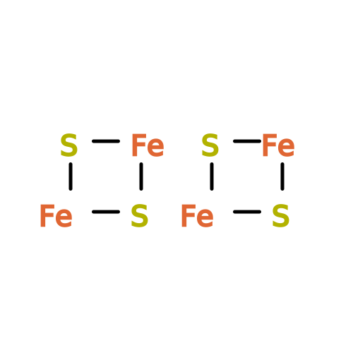 Mass: 351.640 Da / Num. of mol.: 1 / Source method: obtained synthetically / Formula: Fe4S4 / Feature type: SUBJECT OF INVESTIGATION Mass: 351.640 Da / Num. of mol.: 1 / Source method: obtained synthetically / Formula: Fe4S4 / Feature type: SUBJECT OF INVESTIGATION#8: Chemical | ChemComp-CL /  Mass: 35.453 Da / Num. of mol.: 5 / Source method: obtained synthetically / Formula: Cl Mass: 35.453 Da / Num. of mol.: 5 / Source method: obtained synthetically / Formula: Cl#9: Chemical | 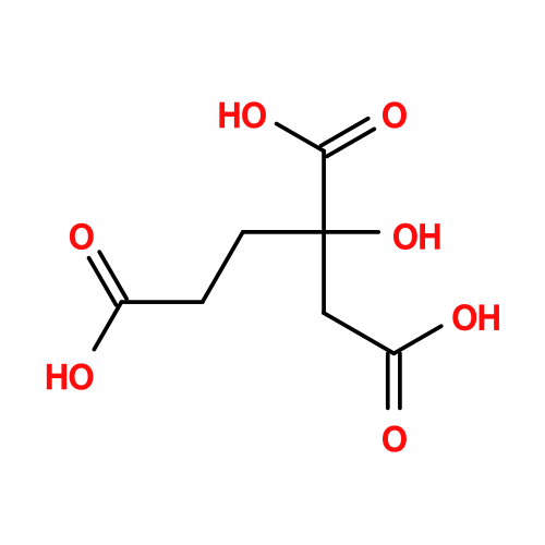 Mass: 206.150 Da / Num. of mol.: 2 / Source method: isolated from a natural source / Formula: C7H10O7 / Feature type: SUBJECT OF INVESTIGATION Mass: 206.150 Da / Num. of mol.: 2 / Source method: isolated from a natural source / Formula: C7H10O7 / Feature type: SUBJECT OF INVESTIGATION#10: Chemical | ChemComp-CLF / | 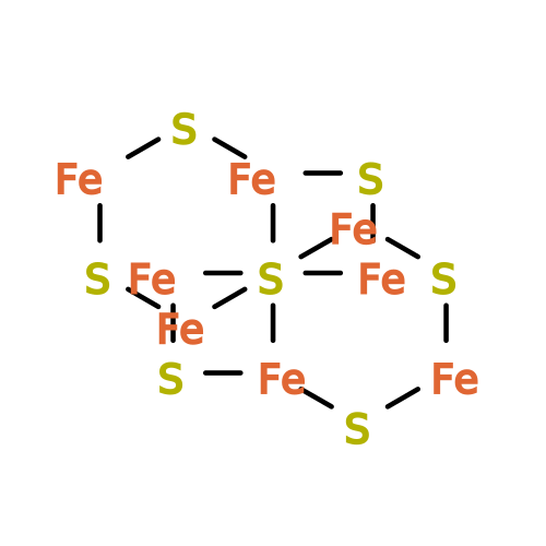 Mass: 671.215 Da / Num. of mol.: 1 / Source method: obtained synthetically / Formula: Fe8S7 / Feature type: SUBJECT OF INVESTIGATION Mass: 671.215 Da / Num. of mol.: 1 / Source method: obtained synthetically / Formula: Fe8S7 / Feature type: SUBJECT OF INVESTIGATION#11: Chemical | ChemComp-F4S / |  Mass: 319.575 Da / Num. of mol.: 1 / Source method: obtained synthetically / Formula: Fe4S3 / Feature type: SUBJECT OF INVESTIGATION Mass: 319.575 Da / Num. of mol.: 1 / Source method: obtained synthetically / Formula: Fe4S3 / Feature type: SUBJECT OF INVESTIGATION#12: Chemical | 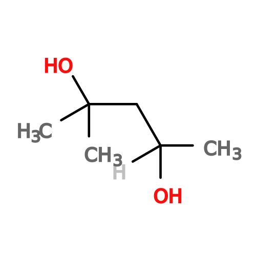 Mass: 118.174 Da / Num. of mol.: 2 / Source method: obtained synthetically / Formula: C6H14O2 / Comment: precipitant*YM Mass: 118.174 Da / Num. of mol.: 2 / Source method: obtained synthetically / Formula: C6H14O2 / Comment: precipitant*YM#13: Chemical |  Mass: 24.305 Da / Num. of mol.: 2 / Source method: obtained synthetically / Formula: Mg / Feature type: SUBJECT OF INVESTIGATION Mass: 24.305 Da / Num. of mol.: 2 / Source method: obtained synthetically / Formula: Mg / Feature type: SUBJECT OF INVESTIGATION#14: Water | ChemComp-HOH / | Mass: 18.015 Da / Num. of mol.: 1454 / Source method: isolated from a natural source / Formula: H2O |
|---|
-Details
| Has ligand of interest | Y |
|---|---|
| Has protein modification | Y |
-Experimental details
-Experiment
| Experiment | Method: X-RAY DIFFRACTION / Number of used crystals: 1 |
|---|
- Sample preparation
| Crystal | Density Matthews: 2.32 Å3/Da / Density % sol: 46.91 % / Description: Thick brown plate |
|---|---|
| Crystal grow | Temperature: 293.15 K / Method: vapor diffusion, sitting drop / pH: 8.5 Details: Samples were centrifuged at 13,000 x g for 3 min to remove macro-aggregates and dust, and crystallised inside an anaerobic chamber (N2/H2 (97:3%) atmosphere, 20 degrees Celsius). ...Details: Samples were centrifuged at 13,000 x g for 3 min to remove macro-aggregates and dust, and crystallised inside an anaerobic chamber (N2/H2 (97:3%) atmosphere, 20 degrees Celsius). Crystallisation was done by the sitting drop method in 96-Well MRC 2-Drop polystyrene Crystallisation Plates (SWISSCI) plate containing 90 uL of crystallisation solution in the reservoir in all cases. Crystals were obtained by mixing 0.7 uL of crystallisation solution with 0.7 uL of protein sample at a concentration of 2.95 mg/mL. The crystallisation solution contained the following: 30 % v/v 2-methyl-2,4-pentanediol, 100 mM Tris pH 8.5, 500 mM Sodium chloride, and 8 % w/v Polyethylene glycol 8,000 (Crystallisation solution of the JBScreen Wizard form Jena Bioscience, Germany). |
-Data collection
| Diffraction | Mean temperature: 100 K / Serial crystal experiment: N |
|---|---|
| Diffraction source | Source: SYNCHROTRON / Site: SLS  / Beamline: X06SA / Wavelength: 1 Å / Beamline: X06SA / Wavelength: 1 Å |
| Detector | Type: DECTRIS EIGER X 16M / Detector: PIXEL / Date: Oct 12, 2022 |
| Radiation | Protocol: SINGLE WAVELENGTH / Monochromatic (M) / Laue (L): M / Scattering type: x-ray |
| Radiation wavelength | Wavelength: 1 Å / Relative weight: 1 |
| Reflection | Resolution: 1.37→78.85 Å / Num. obs: 273586 / % possible obs: 95.3 % / Redundancy: 10.4 % / CC1/2: 0.998 / Rmerge(I) obs: 0.091 / Rpim(I) all: 0.029 / Rrim(I) all: 0.096 / Net I/σ(I): 12.3 |
| Reflection shell | Resolution: 1.37→1.515 Å / Redundancy: 9.6 % / Rmerge(I) obs: 1.198 / Mean I/σ(I) obs: 1.9 / Num. unique obs: 13675 / CC1/2: 0.658 / Rpim(I) all: 0.404 / Rrim(I) all: 1.266 / % possible all: 68.2 |
- Processing
| Software |
| |||||||||||||||||||||||||||||||||||||||||||||||||||||||||||||||||||||||||||||||||||||||||||||||||||||||||||||||||||||||||||||||||||||||||||||||||||||||||||||||||||||||||||||||||||||||||||||||||||||||||||||||||||||||||
|---|---|---|---|---|---|---|---|---|---|---|---|---|---|---|---|---|---|---|---|---|---|---|---|---|---|---|---|---|---|---|---|---|---|---|---|---|---|---|---|---|---|---|---|---|---|---|---|---|---|---|---|---|---|---|---|---|---|---|---|---|---|---|---|---|---|---|---|---|---|---|---|---|---|---|---|---|---|---|---|---|---|---|---|---|---|---|---|---|---|---|---|---|---|---|---|---|---|---|---|---|---|---|---|---|---|---|---|---|---|---|---|---|---|---|---|---|---|---|---|---|---|---|---|---|---|---|---|---|---|---|---|---|---|---|---|---|---|---|---|---|---|---|---|---|---|---|---|---|---|---|---|---|---|---|---|---|---|---|---|---|---|---|---|---|---|---|---|---|---|---|---|---|---|---|---|---|---|---|---|---|---|---|---|---|---|---|---|---|---|---|---|---|---|---|---|---|---|---|---|---|---|---|---|---|---|---|---|---|---|---|---|---|---|---|---|---|---|---|
| Refinement | Method to determine structure: MOLECULAR REPLACEMENT / Resolution: 1.37→21.91 Å / SU ML: 0.1 / Cross valid method: FREE R-VALUE / σ(F): 1.34 / Phase error: 19.51 / Stereochemistry target values: ML Details: The model was manually built via Coot and refined with phenix.refine. The refinement steps were performed by considering all atoms as anisotropic with hydrogens in the riding position. The ...Details: The model was manually built via Coot and refined with phenix.refine. The refinement steps were performed by considering all atoms as anisotropic with hydrogens in the riding position. The model was validated by the MolProbity server.
| |||||||||||||||||||||||||||||||||||||||||||||||||||||||||||||||||||||||||||||||||||||||||||||||||||||||||||||||||||||||||||||||||||||||||||||||||||||||||||||||||||||||||||||||||||||||||||||||||||||||||||||||||||||||||
| Solvent computation | Shrinkage radii: 0.9 Å / VDW probe radii: 1.1 Å / Solvent model: FLAT BULK SOLVENT MODEL | |||||||||||||||||||||||||||||||||||||||||||||||||||||||||||||||||||||||||||||||||||||||||||||||||||||||||||||||||||||||||||||||||||||||||||||||||||||||||||||||||||||||||||||||||||||||||||||||||||||||||||||||||||||||||
| Refinement step | Cycle: LAST / Resolution: 1.37→21.91 Å
| |||||||||||||||||||||||||||||||||||||||||||||||||||||||||||||||||||||||||||||||||||||||||||||||||||||||||||||||||||||||||||||||||||||||||||||||||||||||||||||||||||||||||||||||||||||||||||||||||||||||||||||||||||||||||
| Refine LS restraints |
| |||||||||||||||||||||||||||||||||||||||||||||||||||||||||||||||||||||||||||||||||||||||||||||||||||||||||||||||||||||||||||||||||||||||||||||||||||||||||||||||||||||||||||||||||||||||||||||||||||||||||||||||||||||||||
| LS refinement shell |
|
