[English] 日本語
Yorodumi
- PDB-9sn8: Crystal structure of anthocyanin-related glutathione transferase ... -

+
Open data


ID or keywords:

Loading...

-
Basic information

Entry
Database: PDB / ID: 9sn8
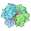
TitleCrystal structure of anthocyanin-related glutathione transferase from bilberry
Componentsglutathione transferase
KeywordsTRANSFERASE / glutathione / glutathione transferase / anthocyanin / cyanidin / bilberry
Biological speciesVaccinium myrtillus (common bilberry)
MethodX-RAY DIFFRACTION / SYNCHROTRON / MOLECULAR REPLACEMENT / Resolution: 2.34 Å
AuthorsDidierjean, C. / Favier, F. / Mathiot, S.
Funding support France, 1items
OrganizationGrant numberCountry
Centre National de la Recherche Scientifique (CNRS) France
CitationJournal: Int.J.Biol.Macromol. / Year: 2026
Title: Structural and biochemical insights into an anthocyanin-related glutathione transferase from bilberry and its inhibition by quercetin.
Authors: Morette, L. / Mathiot, S. / Rochoux, S. / Schwander, T. / Schwartz, M. / Nguyen, H.M. / Favier, F. / Buller, R. / Hecker, A. / Didierjean, C.
History
DepositionSep 10, 2025Deposition site: PDBE / Processing site: PDBE
Revision 1.0Jul 29, 2026Provider: repository / Type: Initial release
Revision 1.1Aug 5, 2026Group: Database references / Category: citation / Item: _citation.journal_volume

-
Structure visualization

Structure viewerMolecule:
MolmilJmol/JSmol

Downloads & links

-
Assembly

Deposited unit
A: glutathione transferase
B: glutathione transferase
C: glutathione transferase
D: glutathione transferase
E: glutathione transferase
F: glutathione transferase
hetero molecules


Theoretical massNumber of molelcules
Total (without water)147,38312
Polymers147,1706
Non-polymers2136
Water4,558253
1
A: glutathione transferase
B: glutathione transferase
hetero molecules


Theoretical massNumber of molelcules
Total (without water)49,1284
Polymers49,0572
Non-polymers712
Water362
TypeNameSymmetry operationNumber
identity operation1_555x,y,z1
Buried area2940 Å2
ΔGint-41 kcal/mol
Surface area19450 Å2
MethodPISA
2
C: glutathione transferase
D: glutathione transferase
hetero molecules


Theoretical massNumber of molelcules
Total (without water)49,1284
Polymers49,0572
Non-polymers712
Water362
TypeNameSymmetry operationNumber
identity operation1_555x,y,z1
Buried area2910 Å2
ΔGint-42 kcal/mol
Surface area19430 Å2
MethodPISA
3
E: glutathione transferase
F: glutathione transferase
hetero molecules


Theoretical massNumber of molelcules
Total (without water)49,1284
Polymers49,0572
Non-polymers712
Water362
TypeNameSymmetry operationNumber
identity operation1_555x,y,z1
Buried area2900 Å2
ΔGint-43 kcal/mol
Surface area19450 Å2
MethodPISA
Unit cell
Length a, b, c (Å)56.294, 159.117, 90.374
Angle α, β, γ (deg.)90, 95.601, 90
Int Tables number4
Space group name H-MP1211
Noncrystallographic symmetry (NCS)NCS domain:
IDEns-IDDetails (eV)
11A
21B
32A
42C
53A
63D
74A
84E
95A
105F
116B
126C
137B
147D
158B
168E
179B
189F
1910C
2010D
2111C
2211E
2312C
2412F
2513D
2613E
2714D
2814F
2915E
3015F

NCS domain segments:

Beg auth comp-ID: VAL / Beg label comp-ID: VAL

Dom-IDComponent-IDEns-IDEnd auth comp-IDEnd label comp-IDAuth asym-IDLabel asym-IDAuth seq-IDLabel seq-ID
111LEULEUAA2 - 2112 - 211
211LEULEUBB2 - 2112 - 211
322METMETAA2 - 2122 - 212
422METMETCC2 - 2122 - 212
533LEULEUAA2 - 2112 - 211
633LEULEUDD2 - 2112 - 211
744METMETAA2 - 2122 - 212
844METMETEE2 - 2122 - 212
955LEULEUAA2 - 2112 - 211
1055LEULEUFF2 - 2112 - 211
1166LEULEUBB2 - 2112 - 211
1266LEULEUCC2 - 2112 - 211
1377ASPASPBB2 - 2132 - 213
1477ASPASPDD2 - 2132 - 213
1588LEULEUBB2 - 2112 - 211
1688LEULEUEE2 - 2112 - 211
1799METMETBB2 - 2122 - 212
1899METMETFF2 - 2122 - 212
191010LEULEUCC2 - 2112 - 211
201010LEULEUDD2 - 2112 - 211
211111METMETCC2 - 2122 - 212
221111METMETEE2 - 2122 - 212
231212LEULEUCC2 - 2112 - 211
241212LEULEUFF2 - 2112 - 211
251313LEULEUDD2 - 2112 - 211
261313LEULEUEE2 - 2112 - 211
271414METMETDD2 - 2122 - 212
281414METMETFF2 - 2122 - 212
291515LEULEUEE2 - 2112 - 211
301515LEULEUFF2 - 2112 - 211

NCS ensembles :
IDDetails (eV)
1Local NCS retraints between domains: 1 2
2Local NCS retraints between domains: 3 4
3Local NCS retraints between domains: 5 6
4Local NCS retraints between domains: 7 8
5Local NCS retraints between domains: 9 10
6Local NCS retraints between domains: 11 12
7Local NCS retraints between domains: 13 14
8Local NCS retraints between domains: 15 16
9Local NCS retraints between domains: 17 18
10Local NCS retraints between domains: 19 20
11Local NCS retraints between domains: 21 22
12Local NCS retraints between domains: 23 24
13Local NCS retraints between domains: 25 26
14Local NCS retraints between domains: 27 28
15Local NCS retraints between domains: 29 30

-
Components

#1: Protein
glutathione transferase


Mass: 24528.400 Da / Num. of mol.: 6
Source method: isolated from a genetically manipulated source
Source: (gene. exp.) Vaccinium myrtillus (common bilberry) / Production host: Escherichia coli (E. coli)
#2: Chemical
ChemComp-CL / CHLORIDE ION


Mass: 35.453 Da / Num. of mol.: 6 / Source method: obtained synthetically / Formula: Cl
#3: Water ChemComp-HOH / water


Mass: 18.015 Da / Num. of mol.: 253 / Source method: isolated from a natural source / Formula: H2O
Has ligand of interestN
Has protein modificationN

-
Experimental details

-
Experiment

ExperimentMethod: X-RAY DIFFRACTION / Number of used crystals: 1

-
Sample preparation

CrystalDensity Matthews: 2.76 Å3/Da / Density % sol: 55.37 %
Crystal growTemperature: 277 K / Method: vapor diffusion
Details: Precipitating solution : - 20% v/v Ethylene glycol / 10 % w/v PEG 8000 - 0,1 M Buffer System 1 pH 6,5 (Buffer System 1 : 1.0M, pH6.5 -> Imidazole; MES monohydrate (acid)) - 0,1 M Amino acids ...Details: Precipitating solution : - 20% v/v Ethylene glycol / 10 % w/v PEG 8000 - 0,1 M Buffer System 1 pH 6,5 (Buffer System 1 : 1.0M, pH6.5 -> Imidazole; MES monohydrate (acid)) - 0,1 M Amino acids (0.2M DL-Glutamic acid monohydrate; 0.2M DL-Alanine; 0.2M Glycine; 0.2M DL-Lysine monohydrochloride; 0.2M DL-Serine) Protein solution : 14.9 mg/mL protein in 20 mM Tris-HCl pH 8.0 ; 200 mM NaCl ; 1mM EDTA

-
Data collection

DiffractionMean temperature: 100 K / Serial crystal experiment: N
Diffraction sourceSource: SYNCHROTRON / Site: ESRF / Beamline: MASSIF-3 / Wavelength: 0.967697 Å
DetectorType: DECTRIS EIGER X 4M / Detector: PIXEL / Date: Nov 11, 2022
RadiationProtocol: SINGLE WAVELENGTH / Monochromatic (M) / Laue (L): M / Scattering type: x-ray
Radiation wavelengthWavelength: 0.967697 Å / Relative weight: 1
ReflectionResolution: 2.34→89.94 Å / Num. obs: 65742 / % possible obs: 98.8 % / Redundancy: 3.5 % / CC1/2: 0.992 / Rmerge(I) obs: 0.105 / Net I/σ(I): 4.8
Reflection shellResolution: 2.34→2.4 Å / Redundancy: 3.7 % / Rmerge(I) obs: 0.637 / Mean I/σ(I) obs: 1.1 / Num. unique obs: 4594 / CC1/2: 0.612 / % possible all: 99

-
Processing

Software
NameVersionClassification
REFMAC5.8.0430 (refmacat 0.4.100)refinement
XDSdata reduction
Aimlessdata scaling
MOLREPphasing
RefinementMethod to determine structure: MOLECULAR REPLACEMENT / Resolution: 2.34→89.94 Å / Cor.coef. Fo:Fc: 0.943 / Cor.coef. Fo:Fc free: 0.924 / SU B: 9.586 / SU ML: 0.213 / Cross valid method: FREE R-VALUE / ESU R: 0.362 / ESU R Free: 0.238 / Details: Hydrogens have not been used
RfactorNum. reflection% reflection
Rfree0.2426 3282 4.995 %
Rwork0.2103 62424 -
all0.212 --
obs-65706 98.72 %
Solvent computationIon probe radii: 0.8 Å / Shrinkage radii: 0.8 Å / VDW probe radii: 1.2 Å / Solvent model: MASK BULK SOLVENT
Displacement parametersBiso mean: 48.692 Å2
Baniso -1Baniso -2Baniso -3
1--0.767 Å2-0 Å2-1.228 Å2
2--0.502 Å20 Å2
3---0.496 Å2
Refinement stepCycle: LAST / Resolution: 2.34→89.94 Å
ProteinNucleic acidLigandSolventTotal
Num. atoms10259 0 6 253 10518
Refine LS restraints
Refine-IDTypeDev idealDev ideal targetNumber
X-RAY DIFFRACTIONr_bond_refined_d0.0070.01210481
X-RAY DIFFRACTIONr_angle_refined_deg1.8541.83914183
X-RAY DIFFRACTIONr_dihedral_angle_1_deg6.33951266
X-RAY DIFFRACTIONr_dihedral_angle_2_deg6.193584
X-RAY DIFFRACTIONr_dihedral_angle_3_deg14.848101897
X-RAY DIFFRACTIONr_dihedral_angle_6_deg14.78810496
X-RAY DIFFRACTIONr_chiral_restr0.1360.21565
X-RAY DIFFRACTIONr_gen_planes_refined0.0080.027950
X-RAY DIFFRACTIONr_nbd_refined0.220.24286
X-RAY DIFFRACTIONr_nbtor_refined0.3050.27140
X-RAY DIFFRACTIONr_xyhbond_nbd_refined0.140.2360
X-RAY DIFFRACTIONr_symmetry_nbd_refined0.2590.299
X-RAY DIFFRACTIONr_symmetry_xyhbond_nbd_refined0.1840.213
X-RAY DIFFRACTIONr_mcbond_it5.764.7465076
X-RAY DIFFRACTIONr_mcangle_it8.8168.526335
X-RAY DIFFRACTIONr_scbond_it7.0764.9745405
X-RAY DIFFRACTIONr_scangle_it10.578.9657847
X-RAY DIFFRACTIONr_lrange_it12.87352.25115288
X-RAY DIFFRACTIONr_ncsr_local_group_10.0760.056779
X-RAY DIFFRACTIONr_ncsr_local_group_20.0760.056854
X-RAY DIFFRACTIONr_ncsr_local_group_30.0760.056827
X-RAY DIFFRACTIONr_ncsr_local_group_40.0790.056842
X-RAY DIFFRACTIONr_ncsr_local_group_50.0790.056791
X-RAY DIFFRACTIONr_ncsr_local_group_60.0760.056795
X-RAY DIFFRACTIONr_ncsr_local_group_70.080.056882
X-RAY DIFFRACTIONr_ncsr_local_group_80.0690.056823
X-RAY DIFFRACTIONr_ncsr_local_group_90.0740.056844
X-RAY DIFFRACTIONr_ncsr_local_group_100.0670.056855
X-RAY DIFFRACTIONr_ncsr_local_group_110.0740.056857
X-RAY DIFFRACTIONr_ncsr_local_group_120.0660.056851
X-RAY DIFFRACTIONr_ncsr_local_group_130.0750.056810
X-RAY DIFFRACTIONr_ncsr_local_group_140.070.056914
X-RAY DIFFRACTIONr_ncsr_local_group_150.0730.056815
Refine LS restraints NCS
Ens-IDDom-IDAuth asym-IDRefine-IDTypeRms dev position (Å)Weight position
11AX-RAY DIFFRACTIONLocal ncs0.076250.0501
12BX-RAY DIFFRACTIONLocal ncs0.076250.0501
23AX-RAY DIFFRACTIONLocal ncs0.076340.0501
24CX-RAY DIFFRACTIONLocal ncs0.076340.0501
35AX-RAY DIFFRACTIONLocal ncs0.075570.0501
36DX-RAY DIFFRACTIONLocal ncs0.075570.0501
47AX-RAY DIFFRACTIONLocal ncs0.078880.05009
48EX-RAY DIFFRACTIONLocal ncs0.078880.05009
59AX-RAY DIFFRACTIONLocal ncs0.07870.05009
510FX-RAY DIFFRACTIONLocal ncs0.07870.05009
611BX-RAY DIFFRACTIONLocal ncs0.076440.05009
612CX-RAY DIFFRACTIONLocal ncs0.076440.05009
713BX-RAY DIFFRACTIONLocal ncs0.079610.0501
714DX-RAY DIFFRACTIONLocal ncs0.079610.0501
815BX-RAY DIFFRACTIONLocal ncs0.069050.05009
816EX-RAY DIFFRACTIONLocal ncs0.069050.05009
917BX-RAY DIFFRACTIONLocal ncs0.074030.05009
918FX-RAY DIFFRACTIONLocal ncs0.074030.05009
1019CX-RAY DIFFRACTIONLocal ncs0.067420.0501
1020DX-RAY DIFFRACTIONLocal ncs0.067420.0501
1121CX-RAY DIFFRACTIONLocal ncs0.073840.05009
1122EX-RAY DIFFRACTIONLocal ncs0.073840.05009
1223CX-RAY DIFFRACTIONLocal ncs0.065530.05009
1224FX-RAY DIFFRACTIONLocal ncs0.065530.05009
1325DX-RAY DIFFRACTIONLocal ncs0.074840.05009
1326EX-RAY DIFFRACTIONLocal ncs0.074840.05009
1427DX-RAY DIFFRACTIONLocal ncs0.070470.05009
1428FX-RAY DIFFRACTIONLocal ncs0.070470.05009
1529EX-RAY DIFFRACTIONLocal ncs0.072580.05009
1530FX-RAY DIFFRACTIONLocal ncs0.072580.05009
LS refinement shell
Resolution (Å)Rfactor RfreeNum. reflection RfreeRfactor RworkNum. reflection RworkRefine-ID% reflection obs (%)
2.34-2.4010.332450.3064572X-RAY DIFFRACTION98.932
2.401-2.4660.3262500.2934504X-RAY DIFFRACTION99.0004
2.466-2.5380.3112300.2734387X-RAY DIFFRACTION99.0348
2.538-2.6160.3082030.2754250X-RAY DIFFRACTION99.1318
2.616-2.7020.2871910.2464215X-RAY DIFFRACTION99.5481
2.702-2.7960.2582140.233972X-RAY DIFFRACTION99.5482
2.796-2.9020.2542370.2253852X-RAY DIFFRACTION99.3199
2.902-3.020.2951820.2143701X-RAY DIFFRACTION99.208
3.02-3.1540.2511760.2043572X-RAY DIFFRACTION98.9702
3.154-3.3080.2341850.2073245X-RAY DIFFRACTION94.1273
3.308-3.4870.251480.2233034X-RAY DIFFRACTION92.7967
3.487-3.6980.2711280.2193111X-RAY DIFFRACTION99.9075
3.698-3.9530.2251500.2022887X-RAY DIFFRACTION99.5411
3.953-4.2690.2191600.172686X-RAY DIFFRACTION99.8246
4.269-4.6750.1991330.1582494X-RAY DIFFRACTION99.6586
4.675-5.2250.2041400.1592228X-RAY DIFFRACTION99.5795
5.225-6.030.236950.211995X-RAY DIFFRACTION99.4291
6.03-7.3770.2411100.2121664X-RAY DIFFRACTION99.8312
7.377-10.3980.203690.1751311X-RAY DIFFRACTION99.639
10.398-89.940.139360.214744X-RAY DIFFRACTION98.8593

+
About Yorodumi

-
News

-
Feb 9, 2022. New format data for meta-information of EMDB entries

New format data for meta-information of EMDB entries

  • Version 3 of the EMDB header file is now the official format.
  • The previous official version 1.9 will be removed from the archive.

Related info.:EMDB header

External links:wwPDB to switch to version 3 of the EMDB data model

-
Aug 12, 2020. Covid-19 info

Covid-19 info

URL: https://pdbj.org/emnavi/covid19.php

New page: Covid-19 featured information page in EM Navigator.

Related info.:Covid-19 info / Mar 5, 2020. Novel coronavirus structure data

+
Mar 5, 2020. Novel coronavirus structure data

Novel coronavirus structure data

Related info.:Yorodumi Speices / Aug 12, 2020. Covid-19 info

External links:COVID-19 featured content - PDBj / Molecule of the Month (242):Coronavirus Proteases

+
Jan 31, 2019. EMDB accession codes are about to change! (news from PDBe EMDB page)

EMDB accession codes are about to change! (news from PDBe EMDB page)

  • The allocation of 4 digits for EMDB accession codes will soon come to an end. Whilst these codes will remain in use, new EMDB accession codes will include an additional digit and will expand incrementally as the available range of codes is exhausted. The current 4-digit format prefixed with “EMD-” (i.e. EMD-XXXX) will advance to a 5-digit format (i.e. EMD-XXXXX), and so on. It is currently estimated that the 4-digit codes will be depleted around Spring 2019, at which point the 5-digit format will come into force.
  • The EM Navigator/Yorodumi systems omit the EMD- prefix.

Related info.:Q: What is EMD? / ID/Accession-code notation in Yorodumi/EM Navigator

External links:EMDB Accession Codes are Changing Soon! / Contact to PDBj

+
Jul 12, 2017. Major update of PDB

Major update of PDB

  • wwPDB released updated PDB data conforming to the new PDBx/mmCIF dictionary.
  • This is a major update changing the version number from 4 to 5, and with Remediation, in which all the entries are updated.
  • In this update, many items about electron microscopy experimental information are reorganized (e.g. em_software).
  • Now, EM Navigator and Yorodumi are based on the updated data.

External links:wwPDB Remediation / Enriched Model Files Conforming to OneDep Data Standards Now Available in the PDB FTP Archive

-
Yorodumi

Thousand views of thousand structures

  • Yorodumi is a browser for structure data from EMDB, PDB, SASBDB, etc.
  • This page is also the successor to EM Navigator detail page, and also detail information page/front-end page for Omokage search.
  • The word "yorodu" (or yorozu) is an old Japanese word meaning "ten thousand". "mi" (miru) is to see.

Related info.:EMDB / PDB / SASBDB / Comparison of 3 databanks / Yorodumi Search / Aug 31, 2016. New EM Navigator & Yorodumi / Yorodumi Papers / Jmol/JSmol / Function and homology information / Changes in new EM Navigator and Yorodumi

Read more