[English] 日本語
Yorodumi
- PDB-9sig: XFEL structure of oxidised Ribonucleotide reductase R2a Y122F mut... -

+
Open data


ID or keywords:

Loading...

-
Basic information

Entry
Database: PDB / ID: 9sig
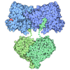
TitleXFEL structure of oxidised Ribonucleotide reductase R2a Y122F mutant from E. coli
ComponentsRibonucleoside-diphosphate reductase 1 subunit beta
KeywordsOXIDOREDUCTASE / Ribonucleotide reductase beta subunit / R2a / Di-iron beta subunit / XFEL structure
Function / homology
Function and homology information


ribonucleoside diphosphate metabolic process / 2'-deoxyribonucleotide biosynthetic process / nucleobase-containing small molecule interconversion / ribonucleoside-diphosphate reductase complex / ribonucleoside-diphosphate reductase / ribonucleoside-diphosphate reductase activity, thioredoxin disulfide as acceptor / deoxyribonucleotide biosynthetic process / iron ion binding / identical protein binding / cytoplasm / cytosol
Similarity search - Function
Ribonucleotide reductase small subunit, acitve site / Ribonucleotide reductase small subunit signature. / Ribonucleotide reductase small subunit / Ribonucleotide reductase small subunit family / Ribonucleotide reductase, small chain / Ribonucleotide reductase-like / Ferritin-like superfamily
Similarity search - Domain/homology
: / Ribonucleoside-diphosphate reductase 1 subunit beta
Similarity search - Component
Biological speciesEscherichia coli (E. coli)
MethodX-RAY DIFFRACTION / FREE ELECTRON LASER / MOLECULAR REPLACEMENT / Resolution: 1.9 Å
AuthorsKumar, R. / Srinivas, V. / Hogbom, M.
Funding support Sweden, United States, 9items
OrganizationGrant numberCountry
Knut and Alice Wallenberg Foundation2023.0201 Sweden
National Institutes of Health/National Institute of General Medical Sciences (NIH/NIGMS)GM149528 United States
National Institutes of Health/National Institute of General Medical Sciences (NIH/NIGMS)GM110501 United States
National Institutes of Health/National Institute of General Medical Sciences (NIH/NIGMS)GM126289 United States
National Institutes of Health/National Institute of General Medical Sciences (NIH/NIGMS)GM117126 United States
National Institutes of Health/National Institute of General Medical Sciences (NIH/NIGMS)GM151988 United States
National Institutes of Health/National Institute of General Medical Sciences (NIH/NIGMS)R24GM154040 United States
Department of Energy (DOE, United States)DE-AC02-76SF00515 United States
Department of Energy (DOE, United States)DE-AC02-05CH11231 United States
CitationJournal: Structure / Year: 2026
Title: Tracking the redox reaction of the iron enzyme ribonucleotide reductase using continuous SerialED and SFX.
Authors: Pacoste, L. / Kumar, R. / Srinivas, V. / Makita, H. / Simon, P.S. / Bannerjee, R. / Minnetian, N.M. / Bhowmick, A. / Paley, D.W. / Mittan-Moreau, D.W. / Chatterjee, K. / Rosenberg, D.J. / ...Authors: Pacoste, L. / Kumar, R. / Srinivas, V. / Makita, H. / Simon, P.S. / Bannerjee, R. / Minnetian, N.M. / Bhowmick, A. / Paley, D.W. / Mittan-Moreau, D.W. / Chatterjee, K. / Rosenberg, D.J. / Batyuk, A. / Gee, L.B. / Alonso-Mori, R. / Sauter, N.K. / Yano, J. / Yachandra, V.K. / John, J. / Aurelius, O. / Brewster, A.S. / Kern, J.F. / Blomberg, B. / Lebrette, H. / Xu, H. / Hofer, G. / Hogbom, M. / Zou, X.
History
DepositionAug 28, 2025Deposition site: PDBE / Processing site: PDBE
Revision 1.0May 13, 2026Provider: repository / Type: Initial release
Revision 1.1May 20, 2026Group: Database references / Category: citation / Item: _citation.pdbx_database_id_PubMed / _citation.title
Revision 1.2Jun 17, 2026Group: Database references / Category: citation / Item: _citation.journal_volume / _citation.page_first

-
Structure visualization

Structure viewerMolecule:
MolmilJmol/JSmol

Downloads & links

-
Assembly

Deposited unit
A: Ribonucleoside-diphosphate reductase 1 subunit beta
B: Ribonucleoside-diphosphate reductase 1 subunit beta
hetero molecules


Theoretical massNumber of molelcules
Total (without water)87,0456
Polymers86,8222
Non-polymers2234
Water4,486249
1


  • Idetical with deposited unit
  • defined by author
  • Evidence: gel filtration
TypeNameSymmetry operationNumber
identity operation1_555x,y,z1
Unit cell
Length a, b, c (Å)73.960, 76.549, 145.738
Angle α, β, γ (deg.)90.000, 90.000, 90.000
Int Tables number19
Space group name H-MP212121
Space group name HallP2ac2ab
Symmetry operation#1: x,y,z
#2: x+1/2,-y+1/2,-z
#3: -x,y+1/2,-z+1/2
#4: -x+1/2,-y,z+1/2

-
Components

#1: Protein Ribonucleoside-diphosphate reductase 1 subunit beta / Protein B2 / Protein R2 / Ribonucleotide reductase 1


Mass: 43410.863 Da / Num. of mol.: 2 / Mutation: Y122F
Source method: isolated from a genetically manipulated source
Source: (gene. exp.) Escherichia coli (E. coli) / Gene: nrdB, ftsB, b2235, JW2229
Production host: Escherichia coli 'BL21-Gold(DE3)pLysS AG' (bacteria)
References: UniProt: P69924, ribonucleoside-diphosphate reductase
#2: Chemical
ChemComp-FE / FE (III) ION


Mass: 55.845 Da / Num. of mol.: 4 / Source method: obtained synthetically / Formula: Fe / Feature type: SUBJECT OF INVESTIGATION
#3: Water ChemComp-HOH / water


Mass: 18.015 Da / Num. of mol.: 249 / Source method: isolated from a natural source / Formula: H2O
Has ligand of interestY
Has protein modificationN

-
Experimental details

-
Experiment

ExperimentMethod: X-RAY DIFFRACTION / Number of used crystals: 1

-
Sample preparation

CrystalDensity Matthews: 2.38 Å3/Da / Density % sol: 48.23 %
Crystal growTemperature: 297 K / Method: batch mode / pH: 5.5 / Details: 0.1M Bis-Tris pH 5.5, PEG 3350 25% / PH range: 5-6 / Temp details: Room Temperature

-
Data collection

DiffractionMean temperature: 297 K / Serial crystal experiment: Y
Diffraction sourceSource: FREE ELECTRON LASER / Site: SLAC LCLS / Beamline: MFX / Wavelength: 1.26295 Å
DetectorType: RAYONIX MX340-HS / Detector: CCD / Date: Jun 12, 2024
RadiationProtocol: SINGLE WAVELENGTH / Monochromatic (M) / Laue (L): M / Scattering type: x-ray
Radiation wavelengthWavelength: 1.26295 Å / Relative weight: 1
ReflectionResolution: 1.9→36.43 Å / Num. obs: 65872 / % possible obs: 99.94 % / Redundancy: 20 % / Biso Wilson estimate: 22.41 Å2 / CC1/2: 0.98 / Net I/σ(I): 5.831
Reflection shellResolution: 1.9→1.93 Å / Num. unique obs: 3197 / CC1/2: 0.41
Serial crystallography sample deliveryMethod: injection

-
Processing

Software
NameVersionClassification
PHENIX1.21.2_5419refinement
cctbx.xfeldata reduction
cxi.mergedata scaling
PHENIXphasing
RefinementMethod to determine structure: MOLECULAR REPLACEMENT / Resolution: 1.9→36.43 Å / SU ML: 0.1757 / Cross valid method: FREE R-VALUE / σ(F): 1.33 / Phase error: 18.4785
Stereochemistry target values: GeoStd + Monomer Library + CDL v1.2
RfactorNum. reflection% reflection
Rfree0.1853 2003 3.04 %
Rwork0.1508 63869 -
obs0.1519 65872 99.94 %
Solvent computationShrinkage radii: 0.9 Å / VDW probe radii: 1.1 Å / Solvent model: FLAT BULK SOLVENT MODEL
Displacement parametersBiso mean: 24.9 Å2
Refinement stepCycle: LAST / Resolution: 1.9→36.43 Å
ProteinNucleic acidLigandSolventTotal
Num. atoms5576 0 4 249 5829
Refine LS restraints
Refine-IDTypeDev idealNumber
X-RAY DIFFRACTIONf_bond_d0.01715746
X-RAY DIFFRACTIONf_angle_d1.34337804
X-RAY DIFFRACTIONf_chiral_restr0.0884864
X-RAY DIFFRACTIONf_plane_restr0.01271003
X-RAY DIFFRACTIONf_dihedral_angle_d17.13082138
LS refinement shell
Resolution (Å)Rfactor RfreeNum. reflection RfreeRfactor RworkNum. reflection RworkRefine-ID% reflection obs (%)
1.9-1.950.30721400.25644452X-RAY DIFFRACTION99.33
1.95-20.27861360.22034524X-RAY DIFFRACTION100
2-2.060.24991480.19754492X-RAY DIFFRACTION100
2.06-2.130.19951390.17184527X-RAY DIFFRACTION99.98
2.13-2.20.20061430.16314494X-RAY DIFFRACTION100
2.2-2.290.20411430.16344496X-RAY DIFFRACTION100
2.29-2.390.19671380.15914571X-RAY DIFFRACTION99.98
2.39-2.520.2311440.164517X-RAY DIFFRACTION99.96
2.52-2.680.19791440.15664540X-RAY DIFFRACTION100
2.68-2.880.19881420.15994576X-RAY DIFFRACTION100
2.88-3.170.18911450.15494582X-RAY DIFFRACTION100
3.17-3.630.17511460.1394613X-RAY DIFFRACTION100
3.63-4.580.12751410.11264644X-RAY DIFFRACTION100
4.58-36.430.14821540.12984841X-RAY DIFFRACTION99.9

+
About Yorodumi

-
News

-
Feb 9, 2022. New format data for meta-information of EMDB entries

New format data for meta-information of EMDB entries

  • Version 3 of the EMDB header file is now the official format.
  • The previous official version 1.9 will be removed from the archive.

Related info.:EMDB header

External links:wwPDB to switch to version 3 of the EMDB data model

-
Aug 12, 2020. Covid-19 info

Covid-19 info

URL: https://pdbj.org/emnavi/covid19.php

New page: Covid-19 featured information page in EM Navigator.

Related info.:Covid-19 info / Mar 5, 2020. Novel coronavirus structure data

+
Mar 5, 2020. Novel coronavirus structure data

Novel coronavirus structure data

Related info.:Yorodumi Speices / Aug 12, 2020. Covid-19 info

External links:COVID-19 featured content - PDBj / Molecule of the Month (242):Coronavirus Proteases

+
Jan 31, 2019. EMDB accession codes are about to change! (news from PDBe EMDB page)

EMDB accession codes are about to change! (news from PDBe EMDB page)

  • The allocation of 4 digits for EMDB accession codes will soon come to an end. Whilst these codes will remain in use, new EMDB accession codes will include an additional digit and will expand incrementally as the available range of codes is exhausted. The current 4-digit format prefixed with “EMD-” (i.e. EMD-XXXX) will advance to a 5-digit format (i.e. EMD-XXXXX), and so on. It is currently estimated that the 4-digit codes will be depleted around Spring 2019, at which point the 5-digit format will come into force.
  • The EM Navigator/Yorodumi systems omit the EMD- prefix.

Related info.:Q: What is EMD? / ID/Accession-code notation in Yorodumi/EM Navigator

External links:EMDB Accession Codes are Changing Soon! / Contact to PDBj

+
Jul 12, 2017. Major update of PDB

Major update of PDB

  • wwPDB released updated PDB data conforming to the new PDBx/mmCIF dictionary.
  • This is a major update changing the version number from 4 to 5, and with Remediation, in which all the entries are updated.
  • In this update, many items about electron microscopy experimental information are reorganized (e.g. em_software).
  • Now, EM Navigator and Yorodumi are based on the updated data.

External links:wwPDB Remediation / Enriched Model Files Conforming to OneDep Data Standards Now Available in the PDB FTP Archive

-
Yorodumi

Thousand views of thousand structures

  • Yorodumi is a browser for structure data from EMDB, PDB, SASBDB, etc.
  • This page is also the successor to EM Navigator detail page, and also detail information page/front-end page for Omokage search.
  • The word "yorodu" (or yorozu) is an old Japanese word meaning "ten thousand". "mi" (miru) is to see.

Related info.:EMDB / PDB / SASBDB / Comparison of 3 databanks / Yorodumi Search / Aug 31, 2016. New EM Navigator & Yorodumi / Yorodumi Papers / Jmol/JSmol / Function and homology information / Changes in new EM Navigator and Yorodumi

Read more