Journal: Protein Sci / Year: 2026 Title: Quaternary structures of Streptococcus pneumoniae nucleoside diphosphate kinase: From hexamers to supramolecular assemblies. Authors: Julie Kerboeuf / Paul Nouri / Frédéric Galisson / Laetitia Daury / Marie-France Giraud / Olivier Lambert / Cédric Orelle / Lionel Ballut / Elise Kaplan / Jean-Michel Jault / Cécile Gonzalez / Abstract: The nucleoside diphosphate kinase (NDK) is a key enzyme that controls the balance of nucleotide pools in all living organisms. Beyond this fundamental role, NDKs exert pleiotropic effects in many ...The nucleoside diphosphate kinase (NDK) is a key enzyme that controls the balance of nucleotide pools in all living organisms. Beyond this fundamental role, NDKs exert pleiotropic effects in many cellular processes, including cell development, signal transduction, differentiation, tumor metastasis, and gene expression. The quaternary structure of NDK is typically hexameric-organized as a trimer of dimers in eukaryotic cells and in many prokaryotes-though in some species it can also be tetrameric. Here, we report the crystal structure of Streptococcus pneumoniae NDK (SpNDK) in its apo state (1.2 Å) and in an adenosine diPhosphate (ADP)-vanadate-bound state (3.4 Å). In both structures, SpNDK adopts a hexameric assembly and the fold of each monomer is highly conserved compared to NDKs from other organisms. A notable feature is the extended Kpn-loop, which plays a key role in stabilizing the hexamer. The protein exhibited remarkably high thermal stability (T ~ 76°C). However, mutation of R28 (R28A), which interacts with the Kpn-loop, destabilized the hexamer, lowering the T by >20°C. Unexpectedly, size-exclusion chromatography and mass photometry revealed that wild-type SpNDK exists as an equilibrium mixture of hexamers, dodecamers, and higher-order supramolecular assemblies. Using cryo-electron microscopy, we solved the three Dimensional (3D) structure of the hexameric state at 2.47 Å resolution, and resolved dodecameric assemblies of the protein. Since the oligomeric state of the NDK influences its cellular function, further investigations will be needed to address the in vivo relevance of these findings, in line with the broad and multifaceted roles of this enzyme family.
Method to determine structure: MOLECULAR REPLACEMENT / Resolution: 3.42→70.396 Å / Cor.coef. Fo:Fc: 0.874 / Cor.coef. Fo:Fc free: 0.834 / SU B: 28.853 / SU ML: 0.431 / Cross valid method: THROUGHOUT / ESU R Free: 0.558 Details: Hydrogens have been added in their riding positions
Rfactor
Num. reflection
% reflection
Selection details
Rfree
0.2688
1027
5.134 %
RANDOM
Rwork
0.204
18978
-
-
all
0.207
-
-
-
obs
-
20005
99.231 %
-
Solvent computation
Ion probe radii: 0.8 Å / Shrinkage radii: 0.8 Å / VDW probe radii: 1.2 Å / Solvent model: MASK BULK SOLVENT
Displacement parameters
Biso mean: 64.535 Å2
Baniso -1
Baniso -2
Baniso -3
1-
2.019 Å2
0 Å2
0 Å2
2-
-
2.019 Å2
-0 Å2
3-
-
-
-4.038 Å2
Refinement step
Cycle: LAST / Resolution: 3.42→70.396 Å
Protein
Nucleic acid
Ligand
Solvent
Total
Num. atoms
6588
0
133
0
6721
Refine LS restraints
Refine-ID
Type
Dev ideal
Dev ideal target
Number
X-RAY DIFFRACTION
r_bond_refined_d
0.004
0.012
6852
X-RAY DIFFRACTION
r_bond_other_d
0.001
0.016
6514
X-RAY DIFFRACTION
r_angle_refined_deg
1.448
1.844
9258
X-RAY DIFFRACTION
r_angle_other_deg
0.489
1.761
15030
X-RAY DIFFRACTION
r_dihedral_angle_1_deg
7.593
5
824
X-RAY DIFFRACTION
r_dihedral_angle_2_deg
8.899
5
54
X-RAY DIFFRACTION
r_dihedral_angle_other_2_deg
0.051
5
4
X-RAY DIFFRACTION
r_dihedral_angle_3_deg
15.945
10
1216
X-RAY DIFFRACTION
r_dihedral_angle_6_deg
13.478
10
320
X-RAY DIFFRACTION
r_chiral_restr
0.066
0.2
1011
X-RAY DIFFRACTION
r_gen_planes_refined
0.004
0.02
7988
X-RAY DIFFRACTION
r_gen_planes_other
0.001
0.02
1560
X-RAY DIFFRACTION
r_nbd_refined
0.216
0.2
1403
X-RAY DIFFRACTION
r_symmetry_nbd_other
0.201
0.2
6088
X-RAY DIFFRACTION
r_nbtor_refined
0.184
0.2
3286
X-RAY DIFFRACTION
r_symmetry_nbtor_other
0.081
0.2
3624
X-RAY DIFFRACTION
r_xyhbond_nbd_refined
0.176
0.2
130
X-RAY DIFFRACTION
r_symmetry_xyhbond_nbd_other
0.039
0.2
3
X-RAY DIFFRACTION
r_metal_ion_refined
0.202
0.2
3
X-RAY DIFFRACTION
r_symmetry_nbd_refined
0.188
0.2
3
X-RAY DIFFRACTION
r_nbd_other
0.186
0.2
24
X-RAY DIFFRACTION
r_symmetry_xyhbond_nbd_refined
0.171
0.2
2
X-RAY DIFFRACTION
r_mcbond_it
4.385
6.21
3314
X-RAY DIFFRACTION
r_mcbond_other
4.384
6.21
3314
X-RAY DIFFRACTION
r_mcangle_it
7.304
11.156
4132
X-RAY DIFFRACTION
r_mcangle_other
7.303
11.157
4133
X-RAY DIFFRACTION
r_scbond_it
4.632
6.81
3538
X-RAY DIFFRACTION
r_scbond_other
4.626
6.81
3535
X-RAY DIFFRACTION
r_scangle_it
7.89
12.336
5126
X-RAY DIFFRACTION
r_scangle_other
7.886
12.333
5124
X-RAY DIFFRACTION
r_lrange_it
11.401
57.375
7434
X-RAY DIFFRACTION
r_lrange_other
11.4
57.396
7435
LS refinement shell
Refine-ID: X-RAY DIFFRACTION / Total num. of bins used: 20
Resolution (Å)
Rfactor Rfree
Num. reflection Rfree
Rfactor Rwork
Num. reflection Rwork
Rfactor all
Num. reflection all
Fsc free
Fsc work
% reflection obs (%)
WRfactor Rwork
3.42-3.509
0.319
77
0.226
1363
0.231
1444
0.925
0.963
99.723
0.214
3.509-3.605
0.33
77
0.316
1337
0.317
1419
0.918
0.916
99.6476
0.307
3.605-3.709
0.561
69
0.516
1281
0.518
1371
0.783
0.838
98.4683
0.522
3.709-3.823
0.329
67
0.329
1264
0.329
1362
0.93
0.931
97.7239
0.322
3.823-3.948
0.332
67
0.219
1209
0.224
1281
0.927
0.966
99.6097
0.21
3.948-4.086
0.266
79
0.185
1203
0.191
1288
0.951
0.975
99.5342
0.177
4.086-4.239
0.257
58
0.16
1168
0.164
1230
0.955
0.982
99.6748
0.158
4.239-4.412
0.209
50
0.156
1111
0.158
1167
0.967
0.983
99.4859
0.156
4.412-4.607
0.214
61
0.133
1073
0.137
1138
0.971
0.988
99.6485
0.134
4.607-4.831
0.21
61
0.121
1033
0.125
1096
0.977
0.99
99.8175
0.125
4.831-5.091
0.193
45
0.123
984
0.126
1032
0.975
0.99
99.7093
0.127
5.091-5.398
0.199
55
0.144
922
0.147
982
0.97
0.989
99.4908
0.146
5.398-5.768
0.226
45
0.15
886
0.153
936
0.971
0.987
99.4658
0.154
5.768-6.227
0.28
41
0.163
819
0.168
873
0.953
0.984
98.5109
0.171
6.227-6.816
0.268
43
0.159
760
0.165
811
0.946
0.982
99.0136
0.171
6.816-7.612
0.211
30
0.148
695
0.151
736
0.972
0.984
98.5054
0.162
7.612-8.772
0.205
40
0.124
623
0.128
667
0.97
0.989
99.4003
0.14
8.772-10.702
0.22
25
0.145
542
0.148
571
0.967
0.987
99.2995
0.167
10.702-14.963
0.172
29
0.182
425
0.181
458
0.985
0.982
99.1266
0.215
14.963-70.396
0.315
8
0.476
280
0.469
295
0.979
0.79
97.6271
1.429
+
About Yorodumi
-
News
-
Feb 9, 2022. New format data for meta-information of EMDB entries
New format data for meta-information of EMDB entries
Version 3 of the EMDB header file is now the official format.
The previous official version 1.9 will be removed from the archive.
In the structure databanks used in Yorodumi, some data are registered as the other names, "COVID-19 virus" and "2019-nCoV". Here are the details of the virus and the list of structure data.
Jan 31, 2019. EMDB accession codes are about to change! (news from PDBe EMDB page)
EMDB accession codes are about to change! (news from PDBe EMDB page)
The allocation of 4 digits for EMDB accession codes will soon come to an end. Whilst these codes will remain in use, new EMDB accession codes will include an additional digit and will expand incrementally as the available range of codes is exhausted. The current 4-digit format prefixed with “EMD-” (i.e. EMD-XXXX) will advance to a 5-digit format (i.e. EMD-XXXXX), and so on. It is currently estimated that the 4-digit codes will be depleted around Spring 2019, at which point the 5-digit format will come into force.
The EM Navigator/Yorodumi systems omit the EMD- prefix.
Related info.:Q: What is EMD? / ID/Accession-code notation in Yorodumi/EM Navigator
Yorodumi is a browser for structure data from EMDB, PDB, SASBDB, etc.
This page is also the successor to EM Navigator detail page, and also detail information page/front-end page for Omokage search.
The word "yorodu" (or yorozu) is an old Japanese word meaning "ten thousand". "mi" (miru) is to see.
Related info.:EMDB / PDB / SASBDB / Comparison of 3 databanks / Yorodumi Search / Aug 31, 2016. New EM Navigator & Yorodumi / Yorodumi Papers / Jmol/JSmol / Function and homology information / Changes in new EM Navigator and Yorodumi
 Movie
Movie Controller

 Yorodumi
 Open data
 Basic information
 Components
 Keywords
 Function and homology information
 Streptococcus pneumoniae R6 (bacteria)
 X-RAY DIFFRACTION /
  Authors
 France, 1items
  Citation
 Structure visualization
 Downloads & links
 Other downloads


 PDBj
PDBj Assembly
 Escherichia coli BL21(DE3) (bacteria) / References: UniProt: P65537, nucleoside-diphosphate kinase
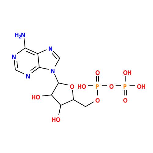 Mass: 427.201 Da / Num. of mol.: 4 / Source method: obtained synthetically / Formula: C10H15N5O10P2 / Feature type: SUBJECT OF INVESTIGATION / Comment: ADP, energy-carrying molecule*YM
 Mass: 98.940 Da / Num. of mol.: 4 / Source method: obtained synthetically / Formula: O3V / Feature type: SUBJECT OF INVESTIGATION
 Mass: 24.305 Da / Num. of mol.: 4 / Source method: obtained synthetically / Formula: Mg / Feature type: SUBJECT OF INVESTIGATION
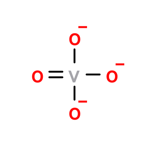 Mass: 114.939 Da / Num. of mol.: 1 / Source method: obtained synthetically / Formula: O4V / Feature type: SUBJECT OF INVESTIGATION
 Sample preparation
 Processing