 Movie
Movie Controller

[English] 日本語
 Yorodumi
- PDB-9s8j: Structure of protein kinase CK2alpha mutant R191Q associated with... -
+ Open data

- Basic information
| Entry | Database: PDB / ID: 9s8j | |||||||||
|---|---|---|---|---|---|---|---|---|---|---|
| Title | Structure of protein kinase CK2alpha mutant R191Q associated with the Okur-Chung Neurodevelopmental Syndrome | |||||||||
 Components Components | Casein kinase II subunit alpha | |||||||||
 Keywords Keywords | TRANSFERASE / Protein kinase CK2 / CK2 / casein kinase II / kinase / Okur-Chung neurodevelopmental syndrome | |||||||||
| Function / homology |  Function and homology information Function and homology informationPhosphorylation and nuclear translocation of BMAL1 (ARNTL) and CLOCK / protein kinase CK2 complex / positive regulation of aggrephagy / regulation of chromosome separation / WNT mediated activation of DVL / Condensation of Prometaphase Chromosomes / symbiont-mediated disruption of host cell PML body / Phosphorylation and nuclear translocation of the CRY:PER:kinase complex / Regulation of CDH1 posttranslational processing and trafficking to plasma membrane / Receptor Mediated Mitophagy ...Phosphorylation and nuclear translocation of BMAL1 (ARNTL) and CLOCK / protein kinase CK2 complex / positive regulation of aggrephagy / regulation of chromosome separation / WNT mediated activation of DVL / Condensation of Prometaphase Chromosomes / symbiont-mediated disruption of host cell PML body / Phosphorylation and nuclear translocation of the CRY:PER:kinase complex / Regulation of CDH1 posttranslational processing and trafficking to plasma membrane / Receptor Mediated Mitophagy / Sin3-type complex / Synthesis of PC / negative regulation of apoptotic signaling pathway / negative regulation of signal transduction by p53 class mediator / Maturation of hRSV A proteins / RUNX1 interacts with co-factors whose precise effect on RUNX1 targets is not known / negative regulation of double-strand break repair via homologous recombination / positive regulation of Wnt signaling pathway / Wnt signaling pathway / negative regulation of proteasomal ubiquitin-dependent protein catabolic process / rhythmic process / Signal transduction by L1 / Hsp90 protein binding / peptidyl-serine phosphorylation / PML body / kinase activity / SPOP-mediated proteasomal degradation of PD-L1(CD274) / Regulation of PTEN stability and activity / double-strand break repair / positive regulation of protein catabolic process / KEAP1-NFE2L2 pathway / positive regulation of cell growth / Cooperation of PDCL (PhLP1) and TRiC/CCT in G-protein beta folding / protein folding / heterochromatin formation / Regulation of TP53 Activity through Phosphorylation / regulation of cell cycle / non-specific serine/threonine protein kinase / protein stabilization / negative regulation of translation / apoptotic process / protein serine kinase activity / protein serine/threonine kinase activity / positive regulation of cell population proliferation / DNA damage response / positive regulation of DNA-templated transcription / chromatin / signal transduction / DNA-templated transcription / nucleoplasm / ATP binding / identical protein binding / nucleus / plasma membrane / cytosol Similarity search - Function | |||||||||
| Biological species |  Homo sapiens (human) Homo sapiens (human) | |||||||||
| Method |  X-RAY DIFFRACTION / SYNCHROTRON / MOLECULAR REPLACEMENT / Resolution: 2.48 Å X-RAY DIFFRACTION / SYNCHROTRON / MOLECULAR REPLACEMENT / Resolution: 2.48 Å | |||||||||
 Authors Authors | Werner, C. / Gast, A. / Meyer, S.C. / Jose, J. / Niefind, K. | |||||||||
| Funding support |  Germany, 2items Germany, 2items
| |||||||||
 Citation Citation | Journal: To Be Published Title: Investigation of the structure-dysfunction relationship of various OCNDS-related CK2alpha mutants Authors: Werner, C. / Gast, A. / Caefer, D. / Fellhoefer, J. / Meyer, S.C. / Jordan, S. / Buchwald, L.M. / Than, T.L. / Schwartz, D. / Jose, J. / Niefind, K. | |||||||||
| History |
|
- Structure visualization
| Structure viewer | Molecule: MolmilJmol/JSmol |
|---|
- Downloads & links
-Download
| PDBx/mmCIF format | 9s8j.cif.gz | 199.6 KB | Display | PDBx/mmCIF format |
|---|---|---|---|---|
| PDB format | pdb9s8j.ent.gz | 125.9 KB | Display | PDB format |
| PDBx/mmJSON format | 9s8j.json.gz | Tree view | PDBx/mmJSON format | |
| Others |  Other downloads Other downloads |
-Validation report
| Arichive directory | https://data.pdbj.org/pub/pdb/validation_reports/s8/9s8jftp://data.pdbj.org/pub/pdb/validation_reports/s8/9s8j | HTTPS FTP |
|---|
-Related structure data


-Links
 PDBj
PDBj


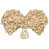

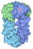
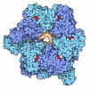
- Assembly
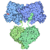
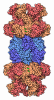
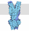
| Deposited unit | 
| |||||||||||||||||||||||||||||||||||||||||||||||||||||||||||||||||||||||||||||||||||||||||||||||||||
|---|---|---|---|---|---|---|---|---|---|---|---|---|---|---|---|---|---|---|---|---|---|---|---|---|---|---|---|---|---|---|---|---|---|---|---|---|---|---|---|---|---|---|---|---|---|---|---|---|---|---|---|---|---|---|---|---|---|---|---|---|---|---|---|---|---|---|---|---|---|---|---|---|---|---|---|---|---|---|---|---|---|---|---|---|---|---|---|---|---|---|---|---|---|---|---|---|---|---|---|---|
| 1 | 
| |||||||||||||||||||||||||||||||||||||||||||||||||||||||||||||||||||||||||||||||||||||||||||||||||||
| 2 | 
| |||||||||||||||||||||||||||||||||||||||||||||||||||||||||||||||||||||||||||||||||||||||||||||||||||
| Unit cell |
| |||||||||||||||||||||||||||||||||||||||||||||||||||||||||||||||||||||||||||||||||||||||||||||||||||
| Noncrystallographic symmetry (NCS) | NCS domain:
NCS domain segments: Ens-ID: ens_1
NCS oper: (Code: givenMatrix: (0.00672020953114, -0.999976928984, -0.000990093161043), (0.997898226331, 0.00664241765458, 0.0644593528748), (-0.0644512891197, -0.00142119256687, 0.997919842243)Vector: ...NCS oper: (Code: given Matrix: (0.00672020953114, -0.999976928984, -0.000990093161043), Vector: |
-Components
| #1: Protein | Mass: 47350.852 Da / Num. of mol.: 2 / Mutation: R191Q Source method: isolated from a genetically manipulated source Source: (gene. exp.) Homo sapiens (human) / Gene: CSNK2A1, CK2A1 / Production host:   Escherichia coli (E. coli) Escherichia coli (E. coli)References: UniProt: P68400, non-specific serine/threonine protein kinase #2: Chemical | 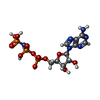 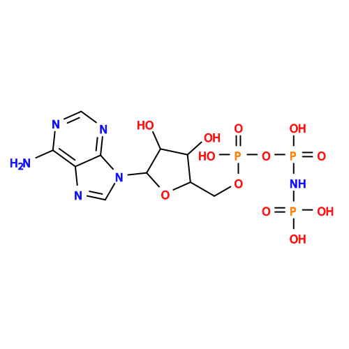 Mass: 506.196 Da / Num. of mol.: 2 / Source method: obtained synthetically / Formula: C10H17N6O12P3 / Comment: AMP-PNP, energy-carrying molecule analogue*YM Mass: 506.196 Da / Num. of mol.: 2 / Source method: obtained synthetically / Formula: C10H17N6O12P3 / Comment: AMP-PNP, energy-carrying molecule analogue*YM#3: Chemical | ChemComp-SO4 /   Mass: 96.063 Da / Num. of mol.: 6 / Source method: obtained synthetically / Formula: SO4 / Feature type: SUBJECT OF INVESTIGATION Mass: 96.063 Da / Num. of mol.: 6 / Source method: obtained synthetically / Formula: SO4 / Feature type: SUBJECT OF INVESTIGATION#4: Chemical | ChemComp-MG /   Mass: 24.305 Da / Num. of mol.: 4 / Source method: obtained synthetically / Formula: Mg Mass: 24.305 Da / Num. of mol.: 4 / Source method: obtained synthetically / Formula: Mg#5: Water | ChemComp-HOH / |  Mass: 18.015 Da / Num. of mol.: 257 / Source method: isolated from a natural source / Formula: H2O Mass: 18.015 Da / Num. of mol.: 257 / Source method: isolated from a natural source / Formula: H2OHas ligand of interest | Y | Has protein modification | N | |
|---|
-Experimental details
-Experiment
| Experiment | Method: X-RAY DIFFRACTION / Number of used crystals: 1 |
|---|
- Sample preparation
| Crystal | Density Matthews: 2.73 Å3/Da / Density % sol: 54.91 % |
|---|---|
| Crystal grow | Temperature: 293 K / Method: vapor diffusion, sitting drop Details: Reservoir: 200 mM Li2SO4, 100 mM Bis-Tris/HCl, pH 6.5, 25 % PEG3350 Protein: 5 mg per mL in 500 mM NaCl, 25 mM Tris/HCl, pH 8.5 Drop: 2 parts protein mixed with 1 part reservoir Soaking with AMPPNP/MgCl2 |
-Data collection
| Diffraction | Mean temperature: 100 K / Serial crystal experiment: N |
|---|---|
| Diffraction source | Source: SYNCHROTRON / Site: ESRF  / Beamline: MASSIF-3 / Wavelength: 0.9677 Å / Beamline: MASSIF-3 / Wavelength: 0.9677 Å |
| Detector | Type: DECTRIS EIGER X 4M / Detector: PIXEL / Date: Apr 30, 2024 |
| Radiation | Protocol: SINGLE WAVELENGTH / Monochromatic (M) / Laue (L): M / Scattering type: x-ray |
| Radiation wavelength | Wavelength: 0.9677 Å / Relative weight: 1 |
| Reflection | Resolution: 2.438→90.708 Å / Num. obs: 28669 / % possible obs: 76.3 % / Redundancy: 8.8 % / Biso Wilson estimate: 19.78 Å2 / CC1/2: 0.979 / Rmerge(I) obs: 0.361 / Net I/σ(I): 6 |
| Reflection shell | Resolution: 2.438→2.722 Å / Rmerge(I) obs: 1.958 / Mean I/σ(I) obs: 1.4 / Num. unique obs: 1433 / CC1/2: 0.461 |
- Processing
| Software |
| |||||||||||||||||||||||||||||||||||||||||||||||||||||||||||||||||||||||||||||||||||||||||||||||||||||||||
|---|---|---|---|---|---|---|---|---|---|---|---|---|---|---|---|---|---|---|---|---|---|---|---|---|---|---|---|---|---|---|---|---|---|---|---|---|---|---|---|---|---|---|---|---|---|---|---|---|---|---|---|---|---|---|---|---|---|---|---|---|---|---|---|---|---|---|---|---|---|---|---|---|---|---|---|---|---|---|---|---|---|---|---|---|---|---|---|---|---|---|---|---|---|---|---|---|---|---|---|---|---|---|---|---|---|---|
| Refinement | Method to determine structure: MOLECULAR REPLACEMENT / Resolution: 2.48→64.14 Å / SU ML: 0.2434 / Cross valid method: FREE R-VALUE / σ(F): 1.33 / Phase error: 26.9775 Stereochemistry target values: GeoStd + Monomer Library + CDL v1.2
| |||||||||||||||||||||||||||||||||||||||||||||||||||||||||||||||||||||||||||||||||||||||||||||||||||||||||
| Solvent computation | Shrinkage radii: 0.9 Å / VDW probe radii: 1.1 Å / Solvent model: FLAT BULK SOLVENT MODEL | |||||||||||||||||||||||||||||||||||||||||||||||||||||||||||||||||||||||||||||||||||||||||||||||||||||||||
| Displacement parameters | Biso mean: 35.62 Å2 | |||||||||||||||||||||||||||||||||||||||||||||||||||||||||||||||||||||||||||||||||||||||||||||||||||||||||
| Refinement step | Cycle: LAST / Resolution: 2.48→64.14 Å
| |||||||||||||||||||||||||||||||||||||||||||||||||||||||||||||||||||||||||||||||||||||||||||||||||||||||||
| Refine LS restraints |
| |||||||||||||||||||||||||||||||||||||||||||||||||||||||||||||||||||||||||||||||||||||||||||||||||||||||||
| Refine LS restraints NCS | Type: Torsion NCS / Rms dev position: 0.756481292814 Å | |||||||||||||||||||||||||||||||||||||||||||||||||||||||||||||||||||||||||||||||||||||||||||||||||||||||||
| LS refinement shell |
|
