 Movie
Movie Controller

[English] 日本語
 Yorodumi
- PDB-9s74: Extracellular serine protease Jep from mouse-adapted S. aureus st... -
+ Open data

- Basic information
| Entry | Database: PDB / ID: 9s74 | ||||||
|---|---|---|---|---|---|---|---|
| Title | Extracellular serine protease Jep from mouse-adapted S. aureus strain JSNZ | ||||||
 Components Components | JSNZ extracellular serine protease Jep | ||||||
 Keywords Keywords | HYDROLASE / serine protease / trypsin-like / beta-barrel / proteolysis / virulence | ||||||
| Function / homology | :  Function and homology information Function and homology information | ||||||
| Biological species |  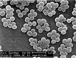 Staphylococcus aureus (bacteria) Staphylococcus aureus (bacteria) | ||||||
| Method |  X-RAY DIFFRACTION / SYNCHROTRON / MOLECULAR REPLACEMENT / Resolution: 0.98 Å X-RAY DIFFRACTION / SYNCHROTRON / MOLECULAR REPLACEMENT / Resolution: 0.98 Å | ||||||
 Authors Authors | Schmoeker, O. / Peringathara, S. / Wolfgramm, H. / Bludau, E. / Girbardt, B. / Palm, G.J. / Hoppen, J. / Holtfreter, S. / Lammers, M. | ||||||
| Funding support |  Germany, 1items Germany, 1items
| ||||||
 Citation Citation | Journal: To Be Published Title: Biochemical and Structural Characterization of novel extracellular serine protease Jep from mouse-adapted S. aureus strain JSNZ Authors: Peringathara, S. / Schmoeker, O. / Bludau, E. / Wolfgramm, H. / Girbardt, B. / Palm, G.J. / Hoppen, J. / Lammers, M. / Holtfreter, S. | ||||||
| History |
|
- Structure visualization
| Structure viewer | Molecule: MolmilJmol/JSmol |
|---|
- Downloads & links
-Download
| PDBx/mmCIF format | 9s74.cif.gz | 209.2 KB | Display | PDBx/mmCIF format |
|---|---|---|---|---|
| PDB format | pdb9s74.ent.gz | 136.8 KB | Display | PDB format |
| PDBx/mmJSON format | 9s74.json.gz | Tree view | PDBx/mmJSON format | |
| Others |  Other downloads Other downloads |
-Validation report
| Arichive directory | https://data.pdbj.org/pub/pdb/validation_reports/s7/9s74ftp://data.pdbj.org/pub/pdb/validation_reports/s7/9s74 | HTTPS FTP |
|---|
-Related structure data

-Links
 PDBj
PDBj- Assembly
| Deposited unit | 
| ||||||||
|---|---|---|---|---|---|---|---|---|---|
| 1 |
| ||||||||
| Unit cell |
| ||||||||
| Components on special symmetry positions |
|
-Components
| #1: Protein | Mass: 23201.006 Da / Num. of mol.: 1 Source method: isolated from a genetically manipulated source Details: The construct corresponds to the the mature protease without N-terminal 35aa signal peptide. Amino acids N59, N60, R61, H62 are not resolved in the crystal structure. The construct carries a ...Details: The construct corresponds to the the mature protease without N-terminal 35aa signal peptide. Amino acids N59, N60, R61, H62 are not resolved in the crystal structure. The construct carries a C-terminal Strep-tag as expression tag (not resolved). Source: (gene. exp.) Staphylococcus aureus (bacteria) / Strain: JSNZ CC88 / Plasmid: pTripleTREP / Details (production host): doi: 10.1186/s12934-025-02736-7Production host: Staphylococcus aureus subsp. aureus RN4220 (bacteria)References: glutamyl endopeptidase | ||||||||
|---|---|---|---|---|---|---|---|---|---|
| #2: Chemical |   Mass: 39.098 Da / Num. of mol.: 2 / Source method: obtained synthetically / Formula: K Mass: 39.098 Da / Num. of mol.: 2 / Source method: obtained synthetically / Formula: K#3: Chemical | ChemComp-SO4 / |  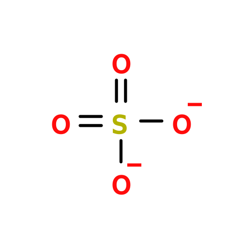 Mass: 96.063 Da / Num. of mol.: 1 / Source method: isolated from a natural source / Formula: SO4 Mass: 96.063 Da / Num. of mol.: 1 / Source method: isolated from a natural source / Formula: SO4#4: Water | ChemComp-HOH / |  Mass: 18.015 Da / Num. of mol.: 247 / Source method: isolated from a natural source / Formula: H2O Mass: 18.015 Da / Num. of mol.: 247 / Source method: isolated from a natural source / Formula: H2OHas ligand of interest | N | Has protein modification | N | |
-Experimental details
-Experiment
| Experiment | Method: X-RAY DIFFRACTION / Number of used crystals: 1 |
|---|
- Sample preparation
| Crystal | Density Matthews: 1.99 Å3/Da / Density % sol: 38.14 % |
|---|---|
| Crystal grow | Temperature: 293 K / Method: vapor diffusion, sitting drop / pH: 6.8 / Details: 0.2M potassium sulfate, 20% PEG 3350 |
-Data collection
| Diffraction | Mean temperature: 100 K / Serial crystal experiment: N | |||||||||||||||||||||||||||
|---|---|---|---|---|---|---|---|---|---|---|---|---|---|---|---|---|---|---|---|---|---|---|---|---|---|---|---|---|
| Diffraction source | Source: SYNCHROTRON / Site: PETRA III, EMBL c/o DESY / Beamline: P13 (MX1) / Wavelength: 0.72932 Å | |||||||||||||||||||||||||||
| Detector | Type: DECTRIS EIGER X 16M / Detector: PIXEL / Date: Dec 1, 2024 | |||||||||||||||||||||||||||
| Radiation | Monochromator: SILICON111 / Protocol: SINGLE WAVELENGTH / Monochromatic (M) / Laue (L): M / Scattering type: x-ray | |||||||||||||||||||||||||||
| Radiation wavelength | Wavelength: 0.72932 Å / Relative weight: 1 | |||||||||||||||||||||||||||
| Reflection | Resolution: 0.98→29.62 Å / Num. obs: 101075 / % possible obs: 97.4 % / Observed criterion σ(I): -3 / Redundancy: 7 % / Biso Wilson estimate: 9.65 Å2 / CC1/2: 0.994 / Rpim(I) all: 0.055 / Rrim(I) all: 0.106 / Rsym value: 0.09 / Χ2: 0.97 / Net I/σ(I): 9.3 | |||||||||||||||||||||||||||
| Reflection shell | Diffraction-ID: 1
|
- Processing
| Software |
| |||||||||||||||||||||||||||||||||||||||||||||||||||||||||||||||||||||||||||||||||||||||||||||||||||||||||||||||||||||||||||||||||||||||||||||||||||||||||||||||||||||
|---|---|---|---|---|---|---|---|---|---|---|---|---|---|---|---|---|---|---|---|---|---|---|---|---|---|---|---|---|---|---|---|---|---|---|---|---|---|---|---|---|---|---|---|---|---|---|---|---|---|---|---|---|---|---|---|---|---|---|---|---|---|---|---|---|---|---|---|---|---|---|---|---|---|---|---|---|---|---|---|---|---|---|---|---|---|---|---|---|---|---|---|---|---|---|---|---|---|---|---|---|---|---|---|---|---|---|---|---|---|---|---|---|---|---|---|---|---|---|---|---|---|---|---|---|---|---|---|---|---|---|---|---|---|---|---|---|---|---|---|---|---|---|---|---|---|---|---|---|---|---|---|---|---|---|---|---|---|---|---|---|---|---|---|---|---|---|
| Refinement | Method to determine structure: MOLECULAR REPLACEMENT / Resolution: 0.98→29.62 Å / Cor.coef. Fo:Fc: 0.975 / Cor.coef. Fo:Fc free: 0.972 / SU B: 0.712 / SU ML: 0.017 / Cross valid method: FREE R-VALUE / ESU R: 0.022 / ESU R Free: 0.022 Details: Hydrogens have been added in their riding positions
| |||||||||||||||||||||||||||||||||||||||||||||||||||||||||||||||||||||||||||||||||||||||||||||||||||||||||||||||||||||||||||||||||||||||||||||||||||||||||||||||||||||
| Solvent computation | Ion probe radii: 0.8 Å / Shrinkage radii: 0.8 Å / VDW probe radii: 1.2 Å / Solvent model: MASK BULK SOLVENT | |||||||||||||||||||||||||||||||||||||||||||||||||||||||||||||||||||||||||||||||||||||||||||||||||||||||||||||||||||||||||||||||||||||||||||||||||||||||||||||||||||||
| Displacement parameters | Biso mean: 16.481 Å2
| |||||||||||||||||||||||||||||||||||||||||||||||||||||||||||||||||||||||||||||||||||||||||||||||||||||||||||||||||||||||||||||||||||||||||||||||||||||||||||||||||||||
| Refinement step | Cycle: LAST / Resolution: 0.98→29.62 Å
| |||||||||||||||||||||||||||||||||||||||||||||||||||||||||||||||||||||||||||||||||||||||||||||||||||||||||||||||||||||||||||||||||||||||||||||||||||||||||||||||||||||
| Refine LS restraints |
| |||||||||||||||||||||||||||||||||||||||||||||||||||||||||||||||||||||||||||||||||||||||||||||||||||||||||||||||||||||||||||||||||||||||||||||||||||||||||||||||||||||
| LS refinement shell |
|
