 Movie
Movie Controller

[English] 日本語
 Yorodumi
- PDB-9s1d: Crystal structure of the methyltransferase ribozyme 1 with two 2'... -
+ Open data

- Basic information
| Entry | Database: PDB / ID: 9s1d | |||||||||
|---|---|---|---|---|---|---|---|---|---|---|
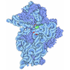
| Title | Crystal structure of the methyltransferase ribozyme 1 with two 2'O-methylation (MTR1m2) | |||||||||
 Components Components |
| |||||||||
 Keywords Keywords | RNA / Ribozyme / methyltransferase / 2'O-methylation / accelerated catalysis | |||||||||
| Function / homology | GUANINE / RNA / RNA (> 10) Function and homology information Function and homology information | |||||||||
| Biological species | synthetic construct (others) | |||||||||
| Method |  X-RAY DIFFRACTION / SYNCHROTRON / MOLECULAR REPLACEMENT / Resolution: 2.6 Å X-RAY DIFFRACTION / SYNCHROTRON / MOLECULAR REPLACEMENT / Resolution: 2.6 Å | |||||||||
 Authors Authors | Chen, H.-A. / Scheitl, C.P.M. / Hoebartner, C. | |||||||||
| Funding support |  Germany, European Union, 2items Germany, European Union, 2items
| |||||||||
 Citation Citation | Journal: Nat Commun / Year: 2026 Title: A magic methyl effect in the active site of a methyltransferase ribozyme. Authors: Aupic, J. / Chen, H.A. / Scheitl, C.P.M. / Hobartner, C. / Magistrato, A. #1: Journal: Nat Chem Biol / Year: 2022Title: Structure and mechanism of the methyltransferase ribozyme MTR1. Authors: Scheitl, C.P.M. / Mieczkowski, M. / Schindelin, H. / Hoebartner, C. | |||||||||
| History |
|
- Structure visualization
| Structure viewer | Molecule: MolmilJmol/JSmol |
|---|
- Downloads & links
-Download
| PDBx/mmCIF format | 9s1d.cif.gz | 114.9 KB | Display | PDBx/mmCIF format |
|---|---|---|---|---|
| PDB format | pdb9s1d.ent.gz | 74.7 KB | Display | PDB format |
| PDBx/mmJSON format | 9s1d.json.gz | Tree view | PDBx/mmJSON format | |
| Others |  Other downloads Other downloads |
-Validation report
| Arichive directory | https://data.pdbj.org/pub/pdb/validation_reports/s1/9s1dftp://data.pdbj.org/pub/pdb/validation_reports/s1/9s1d | HTTPS FTP |
|---|
-Related structure data
| Similar structure data |
|---|
-Links
 PDBj
PDBj
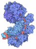
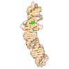
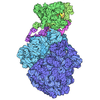
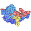
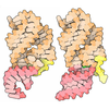
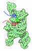

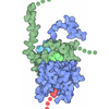
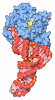
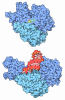
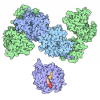

- Assembly
| Deposited unit | 
| ||||||||||||
|---|---|---|---|---|---|---|---|---|---|---|---|---|---|
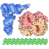
| 1 |
| ||||||||||||
| Unit cell |
|
-Components
-RNA chain , 3 types, 3 molecules ABC
| #1: RNA chain | Mass: 4436.710 Da / Num. of mol.: 1 / Source method: obtained synthetically Details: Chemically synthesized RNA oligonucleotide. N1-methylation of A7 occurred during crystallization process. Source: (synth.) synthetic construct (others) |
|---|---|
| #2: RNA chain | Mass: 7655.662 Da / Num. of mol.: 1 / Source method: obtained synthetically Details: Chemically synthesized RNA oligonucleotide. 2'O-methyl modification at position 12 was introduced during solid-phase synthesis. Source: (synth.) synthetic construct (others) |
| #3: RNA chain | Mass: 7769.729 Da / Num. of mol.: 1 / Source method: obtained synthetically Details: Chemically synthesized RNA oligonucleotide. 2'O-methyl modification at position 42 was introduced during solid-phase synthesis. Source: (synth.) synthetic construct (others) |
-Non-polymers , 4 types, 50 molecules 

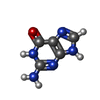

| #4: Chemical |  Mass: 24.305 Da / Num. of mol.: 2 / Source method: obtained synthetically / Formula: Mg Mass: 24.305 Da / Num. of mol.: 2 / Source method: obtained synthetically / Formula: Mg#5: Chemical |  Mass: 22.990 Da / Num. of mol.: 2 / Source method: obtained synthetically / Formula: Na Mass: 22.990 Da / Num. of mol.: 2 / Source method: obtained synthetically / Formula: Na#6: Chemical | ChemComp-GUN / | 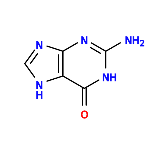 Mass: 151.126 Da / Num. of mol.: 1 / Source method: obtained synthetically / Formula: C5H5N5O / Feature type: SUBJECT OF INVESTIGATION Mass: 151.126 Da / Num. of mol.: 1 / Source method: obtained synthetically / Formula: C5H5N5O / Feature type: SUBJECT OF INVESTIGATION#7: Water | ChemComp-HOH / | Mass: 18.015 Da / Num. of mol.: 45 / Source method: isolated from a natural source / Formula: H2O |
|---|
-Details
| Has ligand of interest | Y |
|---|---|
| Has protein modification | N |
-Experimental details
-Experiment
| Experiment | Method: X-RAY DIFFRACTION / Number of used crystals: 1 |
|---|
- Sample preparation
| Crystal | Density Matthews: 2.53 Å3/Da / Density % sol: 67.16 % / Description: 50-150 um short rod |
|---|---|
| Crystal grow | Temperature: 293.15 K / Method: vapor diffusion, hanging drop / pH: 7.5 Details: 200 uM RNA in a ratio of A:B:C=1:1:1 in 10 mM HEPES pH7.5, 50 mM potassium chloride, 5 mM magnesium chloride and 240 uM of O6-methylguanine Crystallization cocktail: 100 mM NaCl, 100 mM ...Details: 200 uM RNA in a ratio of A:B:C=1:1:1 in 10 mM HEPES pH7.5, 50 mM potassium chloride, 5 mM magnesium chloride and 240 uM of O6-methylguanine Crystallization cocktail: 100 mM NaCl, 100 mM LiCl, 10 mM MgCl2, 50 mM MES pH 6.4-6.7, 36-42% MPD PH range: 6.4-6.7 / Temp details: Growth in a thermally controlled cabinet |
-Data collection
| Diffraction | Mean temperature: 100 K / Ambient temp details: cold nitrogen stream / Serial crystal experiment: N |
|---|---|
| Diffraction source | Source: SYNCHROTRON / Site: PETRA III, DESY / Beamline: P11 / Wavelength: 1.0332 Å |
| Detector | Type: DECTRIS EIGER2 X 16M / Detector: PIXEL / Date: Jun 6, 2021 |
| Radiation | Monochromator: Si-111 and Si-113 reflection / Protocol: SINGLE WAVELENGTH / Monochromatic (M) / Laue (L): M / Scattering type: x-ray |
| Radiation wavelength | Wavelength: 1.0332 Å / Relative weight: 1 |
| Reflection | Resolution: 2.6→43.17 Å / Num. obs: 7084 / % possible obs: 100 % / Observed criterion σ(I): 3 / Redundancy: 25.3 % / Biso Wilson estimate: 76.8 Å2 / CC1/2: 1 / Rmerge(I) obs: 0.044 / Rpim(I) all: 0.01 / Rrim(I) all: 0.045 / Χ2: 1.04 / Net I/σ(I): 42.9 |
| Reflection shell | Resolution: 2.6→2.72 Å / Redundancy: 27 % / Rmerge(I) obs: 2.017 / Mean I/σ(I) obs: 2 / Num. unique obs: 837 / CC1/2: 0.583 / Rpim(I) all: 0.396 / Rrim(I) all: 2.039 / Χ2: 0.82 / % possible all: 100 |
- Processing
| Software |
| ||||||||||||||||||||||||||||||||||||||||||||||||||||||||||||||||||||||||||||||||||||||||||||||||||||
|---|---|---|---|---|---|---|---|---|---|---|---|---|---|---|---|---|---|---|---|---|---|---|---|---|---|---|---|---|---|---|---|---|---|---|---|---|---|---|---|---|---|---|---|---|---|---|---|---|---|---|---|---|---|---|---|---|---|---|---|---|---|---|---|---|---|---|---|---|---|---|---|---|---|---|---|---|---|---|---|---|---|---|---|---|---|---|---|---|---|---|---|---|---|---|---|---|---|---|---|---|---|
| Refinement | Method to determine structure: MOLECULAR REPLACEMENT / Resolution: 2.6→43.17 Å / SU ML: 0.3643 / Cross valid method: FREE R-VALUE / σ(F): 1.37 / Phase error: 26.8536 Stereochemistry target values: GeoStd + Monomer Library + CDL v1.2 Details: Hendrickson-Lattman coefficients were generated in Phenix Resolve based on a non-methylated 7Q7X model, which was used in Phenix Refine as phase restraints (target function: MLHL). Three ...Details: Hendrickson-Lattman coefficients were generated in Phenix Resolve based on a non-methylated 7Q7X model, which was used in Phenix Refine as phase restraints (target function: MLHL). Three methyl groups were introduced during following refinements according to the difference map peaks.
| ||||||||||||||||||||||||||||||||||||||||||||||||||||||||||||||||||||||||||||||||||||||||||||||||||||
| Solvent computation | Shrinkage radii: 0.9 Å / VDW probe radii: 1.1 Å / Solvent model: FLAT BULK SOLVENT MODEL | ||||||||||||||||||||||||||||||||||||||||||||||||||||||||||||||||||||||||||||||||||||||||||||||||||||
| Displacement parameters | Biso mean: 44.34 Å2 | ||||||||||||||||||||||||||||||||||||||||||||||||||||||||||||||||||||||||||||||||||||||||||||||||||||
| Refinement step | Cycle: LAST / Resolution: 2.6→43.17 Å
| ||||||||||||||||||||||||||||||||||||||||||||||||||||||||||||||||||||||||||||||||||||||||||||||||||||
| Refine LS restraints |
| ||||||||||||||||||||||||||||||||||||||||||||||||||||||||||||||||||||||||||||||||||||||||||||||||||||
| LS refinement shell |
| ||||||||||||||||||||||||||||||||||||||||||||||||||||||||||||||||||||||||||||||||||||||||||||||||||||
| Refinement TLS params. | Method: refined / Refine-ID: X-RAY DIFFRACTION
| ||||||||||||||||||||||||||||||||||||||||||||||||||||||||||||||||||||||||||||||||||||||||||||||||||||
| Refinement TLS group |
|
