 Movie
Movie Controller


+ Open data

- Basic information
| Entry | Database: PDB / ID: 9s0w | ||||||
|---|---|---|---|---|---|---|---|
| Title | Crystal structure of Aurora-A bound to DBL3 | ||||||
 Components Components |
| ||||||
 Keywords Keywords | TRANSFERASE / Kinase / Complex / Designed / Inhibitor | ||||||
| Function / homology |  Function and homology information Function and homology informationInteraction between PHLDA1 and AURKA / regulation of centrosome cycle / cilium disassembly / spindle pole centrosome / mitotic centrosome separation / chromosome passenger complex / regulation of G2/M transition of mitotic cell cycle / meiotic spindle organization / spindle organization / positive regulation of mitochondrial fission ...Interaction between PHLDA1 and AURKA / regulation of centrosome cycle / cilium disassembly / spindle pole centrosome / mitotic centrosome separation / chromosome passenger complex / regulation of G2/M transition of mitotic cell cycle / meiotic spindle organization / spindle organization / positive regulation of mitochondrial fission / mitotic spindle pole / spindle midzone / SUMOylation of DNA replication proteins / negative regulation of protein binding / liver regeneration / positive regulation of mitotic cell cycle / positive regulation of mitotic nuclear division / protein serine/threonine/tyrosine kinase activity / centriole / TP53 Regulates Transcription of Genes Involved in G2 Cell Cycle Arrest / AURKA Activation by TPX2 / molecular function activator activity / regulation of signal transduction by p53 class mediator / mitotic spindle organization / regulation of cytokinesis / G2/M transition of mitotic cell cycle / regulation of protein stability / response to wounding / peptidyl-serine phosphorylation / APC/C:Cdh1 mediated degradation of Cdc20 and other APC/C:Cdh1 targeted proteins in late mitosis/early G1 / FBXL7 down-regulates AURKA during mitotic entry and in early mitosis / kinetochore / spindle / microtubule cytoskeleton / mitotic cell cycle / Regulation of PLK1 Activity at G2/M Transition / protein autophosphorylation / spindle pole / ciliary basal body / midbody / Regulation of TP53 Activity through Phosphorylation / basolateral plasma membrane / protein kinase activity / protein phosphorylation / non-specific serine/threonine protein kinase / neuron projection / protein heterodimerization activity / negative regulation of gene expression / protein serine kinase activity / protein serine/threonine kinase activity / centrosome / protein kinase binding / perinuclear region of cytoplasm / nucleoplasm / ATP binding / nucleus / cytosol Similarity search - Function | ||||||
| Biological species |  Homo sapiens (human) Homo sapiens (human)synthetic construct (others) | ||||||
| Method |  X-RAY DIFFRACTION / SYNCHROTRON / MOLECULAR REPLACEMENT / Resolution: 2.07 Å X-RAY DIFFRACTION / SYNCHROTRON / MOLECULAR REPLACEMENT / Resolution: 2.07 Å | ||||||
 Authors Authors | Miles, J.A. / Bayliss, R.W. | ||||||
| Funding support |  United Kingdom, 1items United Kingdom, 1items
| ||||||
 Citation Citation | Journal: Biorxiv / Year: 2026 Title: Selective miniprotein inhibitors of Aurora-A kinase designed using interaction-motif scaffolding Authors: Miles, J.A. / Schiffrin, B. / Holder, J. / Wallis, E.J. / Manfield, I.W. / Burnap, S.A. / Struwe, W.B. / Gergely, F. / Bayliss, R. | ||||||
| History |
|
- Structure visualization
| Structure viewer | Molecule: MolmilJmol/JSmol |
|---|
- Downloads & links
-Download
| PDBx/mmCIF format | 9s0w.cif.gz | 179.4 KB | Display | PDBx/mmCIF format |
|---|---|---|---|---|
| PDB format | pdb9s0w.ent.gz | 116.5 KB | Display | PDB format |
| PDBx/mmJSON format | 9s0w.json.gz | Tree view | PDBx/mmJSON format | |
| Others |  Other downloads Other downloads |
-Validation report
| Arichive directory | https://data.pdbj.org/pub/pdb/validation_reports/s0/9s0wftp://data.pdbj.org/pub/pdb/validation_reports/s0/9s0w | HTTPS FTP |
|---|
-Related structure data
| Related structure data |  9rvkC  9s0kC  9s14C  9s1gC  9s61C  9s7tC  9sq7C  9sv0C C: citing same article ( |
|---|---|
| Similar structure data |
-Links
 PDBj
PDBj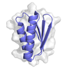








- Assembly
| Deposited unit | 
| ||||||||
|---|---|---|---|---|---|---|---|---|---|
| 1 |
| ||||||||
| Unit cell |
|
-Components
| #1: Protein | Mass: 30874.465 Da / Num. of mol.: 1 Source method: isolated from a genetically manipulated source Source: (gene. exp.) Homo sapiens (human)Gene: AURKA, AIK, AIRK1, ARK1, AURA, AYK1, BTAK, IAK1, STK15, STK6 Production host:   Escherichia coli (E. coli) Escherichia coli (E. coli)References: UniProt: O14965, non-specific serine/threonine protein kinase |
|---|---|
| #2: Protein | Mass: 8044.423 Da / Num. of mol.: 1 Source method: isolated from a genetically manipulated source Source: (gene. exp.) synthetic construct (others) / Production host: Escherichia coli (E. coli) |
| #3: Chemical | ChemComp-ADP /  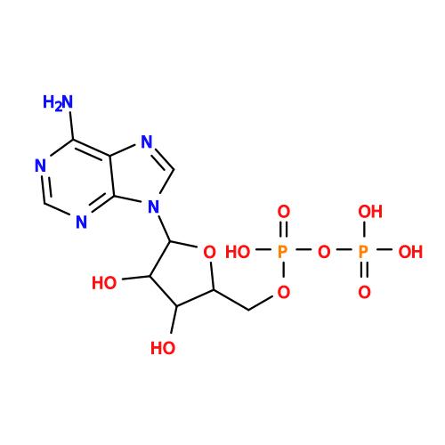 Mass: 427.201 Da / Num. of mol.: 1 / Source method: obtained synthetically / Formula: C10H15N5O10P2 / Comment: ADP, energy-carrying molecule*YM Mass: 427.201 Da / Num. of mol.: 1 / Source method: obtained synthetically / Formula: C10H15N5O10P2 / Comment: ADP, energy-carrying molecule*YM |
| #4: Water | ChemComp-HOH /  Mass: 18.015 Da / Num. of mol.: 48 / Source method: isolated from a natural source / Formula: H2O Mass: 18.015 Da / Num. of mol.: 48 / Source method: isolated from a natural source / Formula: H2O |
| Has ligand of interest | N |
| Has protein modification | N |
-Experimental details
-Experiment
| Experiment | Method: X-RAY DIFFRACTION / Number of used crystals: 1 |
|---|
- Sample preparation
| Crystal | Density Matthews: 2.66 Å3/Da / Density % sol: 53.76 % |
|---|---|
| Crystal grow | Temperature: 291 K / Method: vapor diffusion, sitting drop Details: 0.09M Halides, 0.12M Ethyleneglycol, 0.1M Buffer system 1, 37.5% Precipitant mix 4 |
-Data collection
| Diffraction | Mean temperature: 100 K / Serial crystal experiment: N |
|---|---|
| Diffraction source | Source: SYNCHROTRON / Site: Diamond / Beamline: I03 / Wavelength: 0.9763 Å |
| Detector | Type: DECTRIS EIGER X 16M / Detector: PIXEL / Date: Jun 26, 2025 |
| Radiation | Protocol: SINGLE WAVELENGTH / Monochromatic (M) / Laue (L): M / Scattering type: x-ray |
| Radiation wavelength | Wavelength: 0.9763 Å / Relative weight: 1 |
| Reflection | Resolution: 2.07→47.49 Å / Num. obs: 25854 / % possible obs: 100 % / Redundancy: 13.3 % / CC1/2: 1 / Net I/σ(I): 14.5 |
| Reflection shell | Resolution: 2.07→2.11 Å / Num. unique obs: 1290 / CC1/2: 0.4 |
- Processing
| Software |
| ||||||||||||||||||||||||||||||||||||||||||||||||||||||||||||||||||||||||||||||||||||||||||||||||||||||||||||||||||||||||||||||||||||||||||||||||||||||
|---|---|---|---|---|---|---|---|---|---|---|---|---|---|---|---|---|---|---|---|---|---|---|---|---|---|---|---|---|---|---|---|---|---|---|---|---|---|---|---|---|---|---|---|---|---|---|---|---|---|---|---|---|---|---|---|---|---|---|---|---|---|---|---|---|---|---|---|---|---|---|---|---|---|---|---|---|---|---|---|---|---|---|---|---|---|---|---|---|---|---|---|---|---|---|---|---|---|---|---|---|---|---|---|---|---|---|---|---|---|---|---|---|---|---|---|---|---|---|---|---|---|---|---|---|---|---|---|---|---|---|---|---|---|---|---|---|---|---|---|---|---|---|---|---|---|---|---|---|---|---|---|
| Refinement | Method to determine structure: MOLECULAR REPLACEMENT / Resolution: 2.07→47.49 Å / Cor.coef. Fo:Fc: 0.962 / Cor.coef. Fo:Fc free: 0.947 / SU B: 19.443 / SU ML: 0.22 / Cross valid method: FREE R-VALUE / ESU R: 0.216 / ESU R Free: 0.188 Details: Hydrogens have been added in their riding positions
| ||||||||||||||||||||||||||||||||||||||||||||||||||||||||||||||||||||||||||||||||||||||||||||||||||||||||||||||||||||||||||||||||||||||||||||||||||||||
| Solvent computation | Ion probe radii: 0.9 Å / Shrinkage radii: 0.9 Å / VDW probe radii: 1.1 Å / Solvent model: MASK BULK SOLVENT | ||||||||||||||||||||||||||||||||||||||||||||||||||||||||||||||||||||||||||||||||||||||||||||||||||||||||||||||||||||||||||||||||||||||||||||||||||||||
| Displacement parameters | Biso mean: 48.405 Å2
| ||||||||||||||||||||||||||||||||||||||||||||||||||||||||||||||||||||||||||||||||||||||||||||||||||||||||||||||||||||||||||||||||||||||||||||||||||||||
| Refinement step | Cycle: LAST / Resolution: 2.07→47.49 Å
| ||||||||||||||||||||||||||||||||||||||||||||||||||||||||||||||||||||||||||||||||||||||||||||||||||||||||||||||||||||||||||||||||||||||||||||||||||||||
| Refine LS restraints |
| ||||||||||||||||||||||||||||||||||||||||||||||||||||||||||||||||||||||||||||||||||||||||||||||||||||||||||||||||||||||||||||||||||||||||||||||||||||||
| LS refinement shell |
| ||||||||||||||||||||||||||||||||||||||||||||||||||||||||||||||||||||||||||||||||||||||||||||||||||||||||||||||||||||||||||||||||||||||||||||||||||||||
| Refinement TLS params. | Method: refined / Refine-ID: X-RAY DIFFRACTION
| ||||||||||||||||||||||||||||||||||||||||||||||||||||||||||||||||||||||||||||||||||||||||||||||||||||||||||||||||||||||||||||||||||||||||||||||||||||||
| Refinement TLS group |
|
