[English] 日本語
Yorodumi
- PDB-9rsf: Macaque CD1d with lipid antigen bound -

+
Open data


ID or keywords:

Loading...

-
Basic information

Entry
Database: PDB / ID: 9rsf
TitleMacaque CD1d with lipid antigen bound
Components
  • Beta-2-microglobulin
  • CD1d molecule
KeywordsIMMUNE SYSTEM / CD1d / lipopeptide / lipid antigen / iNKT
Function / homology
Function and homology information


endogenous lipid antigen binding / exogenous lipid antigen binding / antigen processing and presentation, endogenous lipid antigen via MHC class Ib / lipopeptide binding / antigen processing and presentation, exogenous lipid antigen via MHC class Ib / positive regulation of T cell proliferation / cell adhesion molecule binding / early endosome lumen / Nef mediated downregulation of MHC class I complex cell surface expression / DAP12 interactions ...endogenous lipid antigen binding / exogenous lipid antigen binding / antigen processing and presentation, endogenous lipid antigen via MHC class Ib / lipopeptide binding / antigen processing and presentation, exogenous lipid antigen via MHC class Ib / positive regulation of T cell proliferation / cell adhesion molecule binding / early endosome lumen / Nef mediated downregulation of MHC class I complex cell surface expression / DAP12 interactions / T cell mediated cytotoxicity / Endosomal/Vacuolar pathway / Antigen Presentation: Folding, assembly and peptide loading of class I MHC / regulation of iron ion transport / cellular response to iron(III) ion / antigen processing and presentation of exogenous protein antigen via MHC class Ib, TAP-dependent / negative regulation of iron ion transport / negative regulation of forebrain neuron differentiation / regulation of erythrocyte differentiation / peptide antigen assembly with MHC class I protein complex / ER to Golgi transport vesicle membrane / response to molecule of bacterial origin / HFE-transferrin receptor complex / MHC class I peptide loading complex / transferrin transport / negative regulation of receptor-mediated endocytosis / cellular response to iron ion / positive regulation of T cell cytokine production / antigen processing and presentation of endogenous peptide antigen via MHC class I / MHC class I protein complex / peptide antigen assembly with MHC class II protein complex / negative regulation of neurogenesis / multicellular organismal-level iron ion homeostasis / cellular response to nicotine / MHC class II protein complex / positive regulation of receptor-mediated endocytosis / positive regulation of T cell mediated cytotoxicity / negative regulation of epithelial cell proliferation / specific granule lumen / antigen processing and presentation of exogenous peptide antigen via MHC class II / positive regulation of immune response / peptide antigen binding / phagocytic vesicle membrane / recycling endosome membrane / positive regulation of T cell activation / Interferon gamma signaling / Immunoregulatory interactions between a Lymphoid and a non-Lymphoid cell / Modulation by Mtb of host immune system / sensory perception of smell / positive regulation of cellular senescence / tertiary granule lumen / MHC class II protein complex binding / T cell differentiation in thymus / DAP12 signaling / late endosome membrane / negative regulation of neuron projection development / protein refolding / ER-Phagosome pathway / early endosome membrane / amyloid fibril formation / protein homotetramerization / intracellular iron ion homeostasis / learning or memory / endosome membrane / immune response / endoplasmic reticulum lumen / Amyloid fiber formation / Golgi membrane / external side of plasma membrane / innate immune response / lysosomal membrane / focal adhesion / Neutrophil degranulation / SARS-CoV-2 activates/modulates innate and adaptive immune responses / structural molecule activity / Golgi apparatus / endoplasmic reticulum / protein homodimerization activity / : / extracellular exosome / extracellular region / membrane / identical protein binding / plasma membrane / cytosol
Similarity search - Function
MHC-I family domain / Beta-2-Microglobulin / : / MHC class I-like antigen recognition-like / MHC class I-like antigen recognition-like superfamily / MHC classes I/II-like antigen recognition protein / : / Immunoglobulin/major histocompatibility complex, conserved site / Immunoglobulins and major histocompatibility complex proteins signature. / Immunoglobulin C-Type ...MHC-I family domain / Beta-2-Microglobulin / : / MHC class I-like antigen recognition-like / MHC class I-like antigen recognition-like superfamily / MHC classes I/II-like antigen recognition protein / : / Immunoglobulin/major histocompatibility complex, conserved site / Immunoglobulins and major histocompatibility complex proteins signature. / Immunoglobulin C-Type / Immunoglobulin C1-set / Immunoglobulin C1-set domain / Ig-like domain profile. / Immunoglobulin-like domain / Immunoglobulin-like domain superfamily / Immunoglobulin-like fold
Similarity search - Domain/homology
Chem-AGH / CD1d molecule / Beta-2-microglobulin
Similarity search - Component
Biological speciesMacaca mulatta (Rhesus monkey)
Homo sapiens (human)
MethodX-RAY DIFFRACTION / SYNCHROTRON / MOLECULAR REPLACEMENT / Resolution: 1.83 Å
AuthorsBurns, D. / Maly, M. / Tews, I. / Mansour, S.
Funding support United Kingdom, 6items
OrganizationGrant numberCountry
University of Southampton United Kingdom
Medical Research Council (MRC, United Kingdom)MR/W007045/1 United Kingdom
Medical Research Council (MRC, United Kingdom)MR/P023754/1 United Kingdom
Medical Research Council (MRC, United Kingdom)MR/W025728/1 United Kingdom
Medical Research Council (MRC, United Kingdom)MR/S024220/1 United Kingdom
Cancer Research UK23562 United Kingdom
CitationJournal: To Be Published
Title: Structural features of human and macaque CD1d: Implications for antigen presentation
Authors: Burns, D. / Look, A. / Turner, S. / Maly, M. / Essex, J.W. / Tews, I. / Mansour, S.
History
DepositionJul 1, 2025Deposition site: PDBE / Processing site: PDBE
Revision 1.0Jul 15, 2026Provider: repository / Type: Initial release

-
Structure visualization

Structure viewerMolecule:
MolmilJmol/JSmol

Downloads & links

-
Assembly

Deposited unit
A: CD1d molecule
B: Beta-2-microglobulin
C: CD1d molecule
D: Beta-2-microglobulin
hetero molecules


Theoretical massNumber of molelcules
Total (without water)93,07116
Polymers90,8144
Non-polymers2,25712
Water19,2761070
1
A: CD1d molecule
B: Beta-2-microglobulin
hetero molecules


Theoretical massNumber of molelcules
Total (without water)46,4877
Polymers45,4072
Non-polymers1,0805
Water362
TypeNameSymmetry operationNumber
identity operation1_555x,y,z1
Buried area5190 Å2
ΔGint-29 kcal/mol
Surface area18960 Å2
MethodPISA
2
C: CD1d molecule
D: Beta-2-microglobulin
hetero molecules


Theoretical massNumber of molelcules
Total (without water)46,5849
Polymers45,4072
Non-polymers1,1787
Water362
TypeNameSymmetry operationNumber
identity operation1_555x,y,z1
Buried area4890 Å2
ΔGint-31 kcal/mol
Surface area19210 Å2
MethodPISA
Unit cell
Length a, b, c (Å)87.0613, 87.0613, 232.805
Angle α, β, γ (deg.)90, 90, 120
Int Tables number154
Space group name H-MP3221
Components on special symmetry positions
IDModelComponents
11C-305-

CL

21A-497-

HOH

31A-566-

HOH

41A-708-

HOH

51C-741-

HOH

-
Components

-
Protein , 2 types, 4 molecules ACBD

#1: Protein CD1d molecule


Mass: 33527.617 Da / Num. of mol.: 2
Source method: isolated from a genetically manipulated source
Source: (gene. exp.) Macaca mulatta (Rhesus monkey) / Gene: CD1D / Production host: Escherichia coli (E. coli) / Strain (production host): Rossetta DE3 / References: UniProt: F6TR81
#2: Protein Beta-2-microglobulin


Mass: 11879.356 Da / Num. of mol.: 2
Source method: isolated from a genetically manipulated source
Source: (gene. exp.) Homo sapiens (human) / Gene: B2M, CDABP0092, HDCMA22P / Production host: Escherichia coli (E. coli) / Strain (production host): Rossetta DE3 / References: UniProt: P61769

-
Sugars , 1 types, 2 molecules

#3: Sugar ChemComp-AGH / N-{(1S,2R,3S)-1-[(ALPHA-D-GALACTOPYRANOSYLOXY)METHYL]-2,3-DIHYDROXYHEPTADECYL}HEXACOSANAMIDE


Type: D-saccharide / Mass: 858.322 Da / Num. of mol.: 2 / Source method: obtained synthetically / Formula: C50H99NO9 / Feature type: SUBJECT OF INVESTIGATION

-
Non-polymers , 3 types, 1080 molecules

#4: Chemical
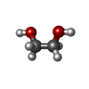
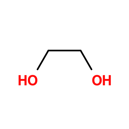
ChemComp-EDO / 1,2-ETHANEDIOL / ETHYLENE GLYCOL


Mass: 62.068 Da / Num. of mol.: 7 / Source method: obtained synthetically / Formula: C2H6O2
#5: Chemical ChemComp-CL / CHLORIDE ION


Mass: 35.453 Da / Num. of mol.: 3 / Source method: obtained synthetically / Formula: Cl
#6: Water ChemComp-HOH / water


Mass: 18.015 Da / Num. of mol.: 1070 / Source method: isolated from a natural source / Formula: H2O

-
Details

Has ligand of interestY
Has protein modificationY

-
Experimental details

-
Experiment

ExperimentMethod: X-RAY DIFFRACTION / Number of used crystals: 1

-
Sample preparation

CrystalDensity Matthews: 2.97 Å3/Da / Density % sol: 58.64 %
Crystal growTemperature: 293.15 K / Method: vapor diffusion, sitting drop / pH: 6.5
Details: 10% w/v PEG 8000, 20% v/v ethylene glycol 0.03 M, 0.1 M MES/imidazole pH 6.5

-
Data collection

DiffractionMean temperature: 100 K / Serial crystal experiment: N
Diffraction sourceSource: SYNCHROTRON / Site: Diamond / Beamline: I03 / Wavelength: 0.9763 Å
DetectorType: DECTRIS EIGER2 XE 16M / Detector: PIXEL / Date: Dec 11, 2022
RadiationProtocol: SINGLE WAVELENGTH / Monochromatic (M) / Laue (L): M / Scattering type: x-ray
Radiation wavelengthWavelength: 0.9763 Å / Relative weight: 1
ReflectionResolution: 1.83→54.08 Å / Num. obs: 89434 / % possible obs: 98.22 % / Redundancy: 17.1 % / Biso Wilson estimate: 24.77 Å2 / CC1/2: 1 / CC star: 1 / Rpim(I) all: 0.013 / Rrim(I) all: 0.056 / Net I/σ(I): 32.7
Reflection shellResolution: 1.83→1.86 Å / Redundancy: 6.7 % / Mean I/σ(I) obs: 2.1 / Num. unique obs: 25554 / CC1/2: 0.914 / CC star: 0.977 / Rpim(I) all: 0.179 / Rrim(I) all: 0.486 / % possible all: 84.73

-
Processing

Software
NameVersionClassification
DIALS3.21data reduction
xia2data scaling
gemmi0.7.1data extraction
MOLREPphasing
Coot0.9.8.86model building
Servalcat0.4.113refinement
RefinementMethod to determine structure: MOLECULAR REPLACEMENT / Resolution: 1.83→54.076 Å / Cross valid method: FREE R-VALUE
RfactorNum. reflection% reflection
Rfree0.2171 2075 -
Rwork0.171 87274 -
obs-89349 98.12 %
Displacement parametersBiso mean: 33.15 Å2
Baniso -1Baniso -2Baniso -3
1-2.81595458 Å20 Å20 Å2
2--2.81595458 Å20 Å2
3----5.63190917 Å2
Refinement stepCycle: LAST / Resolution: 1.83→54.076 Å
ProteinNucleic acidLigandSolventTotal
Num. atoms6056 0 151 1070 7277
Refine LS restraints
Refine-IDTypeDev idealNumberWeight
X-RAY DIFFRACTIONs_bond_nonh_d0.013765600.0119
X-RAY DIFFRACTIONs_angle_nonh_deg2.033589271.8143
X-RAY DIFFRACTIONs_dihedral_angle_1_deg7.22258015
X-RAY DIFFRACTIONs_dihedral_angle_2_deg3.03352145
X-RAY DIFFRACTIONs_dihedral_angle_3_deg12.2411215810
X-RAY DIFFRACTIONs_dihedral_angle_6_deg15.277438910
X-RAY DIFFRACTIONs_chiral_restr0.09779330.132
X-RAY DIFFRACTIONs_planes0.015394560.02
X-RAY DIFFRACTIONs_nbd0.214192710.2
X-RAY DIFFRACTIONs_nbtor0.2246109530.2
X-RAY DIFFRACTIONs_hbond_nbd0.16718930.2
X-RAY DIFFRACTIONs_symmetry_nbd0.19281430.2
X-RAY DIFFRACTIONs_symmetry_hbond_nbd0.1974670.2
LS refinement shell

Refine-ID: X-RAY DIFFRACTION

Resolution (Å)Rfactor RfreeNum. reflection RfreeRfactor RworkNum. reflection RworkCor.coef. Fo:FcCor.coef. Fo:Fc freeNum. reflection obs% reflection obs (%)
1.83-1.8650.2498780.237640840.88450.8452416284.8
1.865-1.9020.26151110.210742600.89390.8522437189.51
1.902-1.9410.29361060.208843470.89310.7583445393.87
1.941-1.9830.2677780.179844640.92420.8806454297.28
1.983-2.0280.18861100.167843820.9290.9035449299.38
2.028-2.0750.20661110.16143640.93680.88634475100
2.075-2.1260.23041070.162242730.93750.88084380100
2.126-2.1810.2960.168641570.9310.89594253100
2.181-2.2410.2548990.167240390.93180.85944138100
2.241-2.3060.1928850.160639410.93670.88614026100
2.306-2.3770.23361020.162438040.9330.9042390699.95
2.377-2.4550.2014870.158937260.93530.91853813100
2.455-2.5410.2093780.159936190.93850.84573697100
2.541-2.6360.2034860.157135110.93850.90513597100
2.636-2.7440.1878860.162733280.93640.90083414100
2.744-2.8650.216850.162831980.93140.90733283100
2.865-3.0050.2012830.168131240.92740.915320799.97
3.005-3.1670.1989600.175729170.91880.86982977100
3.167-3.3580.2457770.176428040.92810.89332881100
3.358-3.5890.2077760.180226190.92840.9052695100
3.589-3.8750.2014390.165924850.93780.8918252499.96
3.875-4.2430.2254590.151822590.94310.85052318100
4.243-4.740.187520.137120750.95260.87332127100
4.74-5.4660.2303370.16518690.93940.92151906100
5.466-6.6780.2291320.211715930.91320.88091625100
6.678-9.3730.2731420.206712490.90610.85931291100
9.373-54.0760.1635130.22177830.87290.9374796100

+
About Yorodumi

-
News

-
Feb 9, 2022. New format data for meta-information of EMDB entries

New format data for meta-information of EMDB entries

  • Version 3 of the EMDB header file is now the official format.
  • The previous official version 1.9 will be removed from the archive.

Related info.:EMDB header

External links:wwPDB to switch to version 3 of the EMDB data model

-
Aug 12, 2020. Covid-19 info

Covid-19 info

URL: https://pdbj.org/emnavi/covid19.php

New page: Covid-19 featured information page in EM Navigator.

Related info.:Covid-19 info / Mar 5, 2020. Novel coronavirus structure data

+
Mar 5, 2020. Novel coronavirus structure data

Novel coronavirus structure data

Related info.:Yorodumi Speices / Aug 12, 2020. Covid-19 info

External links:COVID-19 featured content - PDBj / Molecule of the Month (242):Coronavirus Proteases

+
Jan 31, 2019. EMDB accession codes are about to change! (news from PDBe EMDB page)

EMDB accession codes are about to change! (news from PDBe EMDB page)

  • The allocation of 4 digits for EMDB accession codes will soon come to an end. Whilst these codes will remain in use, new EMDB accession codes will include an additional digit and will expand incrementally as the available range of codes is exhausted. The current 4-digit format prefixed with “EMD-” (i.e. EMD-XXXX) will advance to a 5-digit format (i.e. EMD-XXXXX), and so on. It is currently estimated that the 4-digit codes will be depleted around Spring 2019, at which point the 5-digit format will come into force.
  • The EM Navigator/Yorodumi systems omit the EMD- prefix.

Related info.:Q: What is EMD? / ID/Accession-code notation in Yorodumi/EM Navigator

External links:EMDB Accession Codes are Changing Soon! / Contact to PDBj

+
Jul 12, 2017. Major update of PDB

Major update of PDB

  • wwPDB released updated PDB data conforming to the new PDBx/mmCIF dictionary.
  • This is a major update changing the version number from 4 to 5, and with Remediation, in which all the entries are updated.
  • In this update, many items about electron microscopy experimental information are reorganized (e.g. em_software).
  • Now, EM Navigator and Yorodumi are based on the updated data.

External links:wwPDB Remediation / Enriched Model Files Conforming to OneDep Data Standards Now Available in the PDB FTP Archive

-
Yorodumi

Thousand views of thousand structures

  • Yorodumi is a browser for structure data from EMDB, PDB, SASBDB, etc.
  • This page is also the successor to EM Navigator detail page, and also detail information page/front-end page for Omokage search.
  • The word "yorodu" (or yorozu) is an old Japanese word meaning "ten thousand". "mi" (miru) is to see.

Related info.:EMDB / PDB / SASBDB / Comparison of 3 databanks / Yorodumi Search / Aug 31, 2016. New EM Navigator & Yorodumi / Yorodumi Papers / Jmol/JSmol / Function and homology information / Changes in new EM Navigator and Yorodumi

Read more