 Movie
Movie Controller

[English] 日本語
 Yorodumi
- PDB-9rpr: Cryo-EM structure of LptDEM complex containing Shigella flexneri ... -
+ Open data

- Basic information
| Entry | Database: PDB / ID: 9rpr | |||||||||
|---|---|---|---|---|---|---|---|---|---|---|
| Title | Cryo-EM structure of LptDEM complex containing Shigella flexneri LptE and endogenous E. coli LptD and LptM | |||||||||
 Components Components |
| |||||||||
 Keywords Keywords | MEMBRANE PROTEIN / lipopolysaccharide transport / outer membrane protein complex / beta-barrel / lipoprotein | |||||||||
| Function / homology |  Function and homology information Function and homology informationtransporter complex / lipopolysaccharide transport / Gram-negative-bacterium-type cell outer membrane assembly / cell outer membrane / lipopolysaccharide binding Similarity search - Function | |||||||||
| Biological species |  Shigella flexneri (bacteria) Shigella flexneri (bacteria) | |||||||||
| Method | ELECTRON MICROSCOPY / single particle reconstruction / cryo EM / Resolution: 2.78 Å | |||||||||
 Authors Authors | Dunbar, E. / Basle, A. / van den Berg, B. | |||||||||
| Funding support |  United Kingdom, 2items United Kingdom, 2items
| |||||||||
 Citation Citation | Journal: Proc Natl Acad Sci U S A / Year: 2025 Title: Small siphophage binding to an open state of the LptDE outer membrane lipopolysaccharide translocon. Authors: Emily Dunbar / Robert Clark / Arnaud Baslé / Shenaz Allyjaun / Hector Newman / Julia Hubbard / Syma Khalid / Bert van den Berg / Abstract: Bacteriophages are bacterial viruses that provide alternatives to small-molecule drugs to combat infections by antibiotic-resistant bacteria. To infect a bacterial host, a phage needs to bind to the ...Bacteriophages are bacterial viruses that provide alternatives to small-molecule drugs to combat infections by antibiotic-resistant bacteria. To infect a bacterial host, a phage needs to bind to the bacterial surface via receptor binding proteins (RBPs), which are critical for determining host specificity. For functionally important receptors, the RBP-receptor interaction could be exploited via phage steering, where emerging bacterial resistance due to receptor modification could make bacteria less fit or virulent. Despite this, relatively little is known about RBP-receptor interactions. Here, we build on the recent discovery of coliphages that have the outer membrane (OM) lipopolysaccharide translocon LptDE as their terminal receptor and show via cryogenic electron microscopy that, surprisingly, the RBP of the small siphophage Oekolampad binds to a hitherto unobserved, open state of LptDE. The open lateral gate of LptD is occupied by a β-strand peptide originating from the degraded N-terminal jellyroll domain of LptD, suggesting the possibility of LptD inhibition via peptidomimetics. A structure of LptDE in complex with the superinfection exclusion (SE) protein Rtp45 of the Oekolampad-related phage Rtp shows a mechanism of SE where Rtp45-induced conformational changes in LptD resulting from steric clashes preclude RBP binding. Finally, analysis of spontaneous Oekolampad-resistant mutants identifies mutations in LptD that abolish the LptDE-RBP interaction in vitro. SDS-EDTA sensitivity assays of the mutants show no major OM defects, consistent with largely preserved LptDE function, and suggesting that phage steering via LptDE might be challenging. | |||||||||
| History |
|
- Structure visualization
| Structure viewer | Molecule: MolmilJmol/JSmol |
|---|
- Downloads & links
-Download
| PDBx/mmCIF format | 9rpr.cif.gz | 196.3 KB | Display | PDBx/mmCIF format |
|---|---|---|---|---|
| PDB format | pdb9rpr.ent.gz | 151.3 KB | Display | PDB format |
| PDBx/mmJSON format | 9rpr.json.gz | Tree view | PDBx/mmJSON format | |
| Others |  Other downloads Other downloads |
-Validation report
| Arichive directory | https://data.pdbj.org/pub/pdb/validation_reports/rp/9rprftp://data.pdbj.org/pub/pdb/validation_reports/rp/9rpr | HTTPS FTP |
|---|
-Related structure data
| Related structure data |  54169MC  9rpsC  9rptC  9rpwC  9rqiC M: map data used to model this data C: citing same article ( |
|---|---|
| Similar structure data |


-Links
 PDBj
PDBj


- Assembly
| Deposited unit | 
|
|---|---|
| 1 |
|
-Components
| #1: Protein | Mass: 22240.500 Da / Num. of mol.: 1 Source method: isolated from a genetically manipulated source Source: (gene. exp.) Shigella flexneri (bacteria) / Gene: lptE, rlpB, SF0640, S0662 / Production host: |
|---|---|
| #2: Protein | Mass: 89754.719 Da / Num. of mol.: 1 / Source method: isolated from a natural source / Source: (natural) |
| #3: Protein/peptide | Mass: 5103.607 Da / Num. of mol.: 1 / Source method: isolated from a natural source / Source: (natural) |
| #4: Chemical | ChemComp-PLM /   Mass: 256.424 Da / Num. of mol.: 1 / Source method: obtained synthetically / Formula: C16H32O2 Mass: 256.424 Da / Num. of mol.: 1 / Source method: obtained synthetically / Formula: C16H32O2 |
| #5: Chemical | ChemComp-PXS / (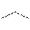  Mass: 552.912 Da / Num. of mol.: 1 / Source method: obtained synthetically / Formula: C35H68O4 Mass: 552.912 Da / Num. of mol.: 1 / Source method: obtained synthetically / Formula: C35H68O4 |
| Has ligand of interest | N |
| Has protein modification | Y |
-Experimental details
-Experiment
| Experiment | Method: ELECTRON MICROSCOPY |
|---|---|
| EM experiment | Aggregation state: PARTICLE / 3D reconstruction method: single particle reconstruction |
- Sample preparation
| Component |
| ||||||||||||||||||||||||||||
|---|---|---|---|---|---|---|---|---|---|---|---|---|---|---|---|---|---|---|---|---|---|---|---|---|---|---|---|---|---|
| Molecular weight |
| ||||||||||||||||||||||||||||
| Source (natural) |
| ||||||||||||||||||||||||||||
| Source (recombinant) |
| ||||||||||||||||||||||||||||
| Buffer solution | pH: 7.5 / Details: 10mM HEPES, pH7.5, 100mM NaCl, 0.05% (w/v) DDM | ||||||||||||||||||||||||||||
| Buffer component |
| ||||||||||||||||||||||||||||
| Specimen | Embedding applied: NO / Shadowing applied: NO / Staining applied: NO / Vitrification applied: YES | ||||||||||||||||||||||||||||
| Specimen support | Grid material: COPPER / Grid mesh size: 200 divisions/in. / Grid type: Quantifoil R1.2/1.3 | ||||||||||||||||||||||||||||
| Vitrification | Cryogen name: ETHANE / Humidity: 100 % / Chamber temperature: 277.15 K |
- Electron microscopy imaging
| Experimental equipment |  Model: Titan Krios / Image courtesy: FEI Company |
|---|---|
| Microscopy | Model: TFS KRIOS |
| Electron gun | Electron source:  FIELD EMISSION GUN / Accelerating voltage: 300 kV / Illumination mode: FLOOD BEAM FIELD EMISSION GUN / Accelerating voltage: 300 kV / Illumination mode: FLOOD BEAM |
| Electron lens | Mode: BRIGHT FIELD / Nominal magnification: 165000 X / Nominal defocus max: 2000 nm / Nominal defocus min: 800 nm / Cs: 2.7 mm / C2 aperture diameter: 50 µm |
| Image recording | Electron dose: 50 e/Å2 / Film or detector model: FEI FALCON IV (4k x 4k) / Num. of grids imaged: 1 / Num. of real images: 7277 |
- Processing
| EM software |
| |||||||||||||||
|---|---|---|---|---|---|---|---|---|---|---|---|---|---|---|---|---|
| CTF correction | Type: NONE | |||||||||||||||
| Particle selection | Num. of particles selected: 1081511 | |||||||||||||||
| 3D reconstruction | Resolution: 2.78 Å / Resolution method: FSC 0.143 CUT-OFF / Num. of particles: 112032 / Symmetry type: POINT | |||||||||||||||
| Atomic model building | Source name: AlphaFold / Type: in silico model |
